Tendon Problems of the Ulnar Wrist
- Affiliations
-
- 1Department of Orthopedic Surgery, Gachon University School of Medicine, Incheon, Korea.
- 2Department of Orthopedic Surgery, Yonsei University Wonju College of Medicine, Wonju, Korea. jroh@yonsei.ac.kr
- KMID: 2438978
- DOI: http://doi.org/10.4055/jkoa.2017.52.2.138
Abstract
- It is challenging for orthopedic surgeons to diagnose pain at the ulnar aspect of the wrist due to the small and complex anatomical structures involved. Ulnar-sided wrist pain can also result from tendon problems, including extensor carpi ulnaris tendon and flexor carpi ulnaris tendon. Disorders of the extensor carpi ulnaris tendon include subluxation, dislocation, stenosing tenosynovitis, and tendinopathy. Unlike the extensor carpi ulnaris tendon which is prone to subluxation, dislocation and stenosing tenosynovitis from passing through as sheath, a flexor carpi ulnaris tendon is unsheathed, and calcific tendinitis and crystal deposition disease can occur at the distal tendinous portion of the flexor carpi ulnaris tendon.
Keyword
Figure
-
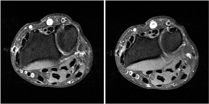
Figure 1 Axial fast spin echo proton density magnetic resonance imaging scan of the wrist. Extensor carpi ulnaris tendinitis is presented as inflammation of the synovial lining of the extensor carpi ulnaris and is frequently associated with intrinsic tendon degeneration.
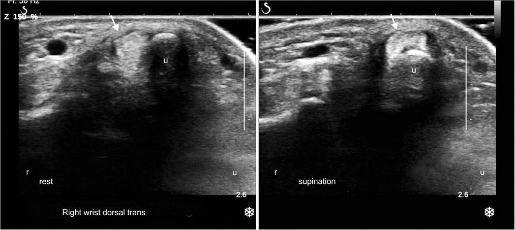
Figure 2 Axial dynamic ultrasound images. In wrist supination, extensor carpi ulnaris (ECU) tendon leaves its sheath or its osseous groove. Left arrow, normal relationship between ECU and ulnar styloid process; right arrow, ECU dislocated on ulnar styloid process; u, ulnar styloid process.
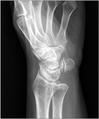
Figure 3 Thirty-degree supinated lateral radiograph demonstrating pisotriquetral arthritis.
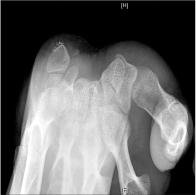
Figure 4 Calcification of the flexor carpi ulnaris just proximal to the insertion on the pisiform is shown in the carpal tunnel view.

Figure 5 Longitudinal ultrasound image of the flexor carpi ulnaris (FCU). Irregular calcifications around FCU tendon insertion site of the pisiform is shown.
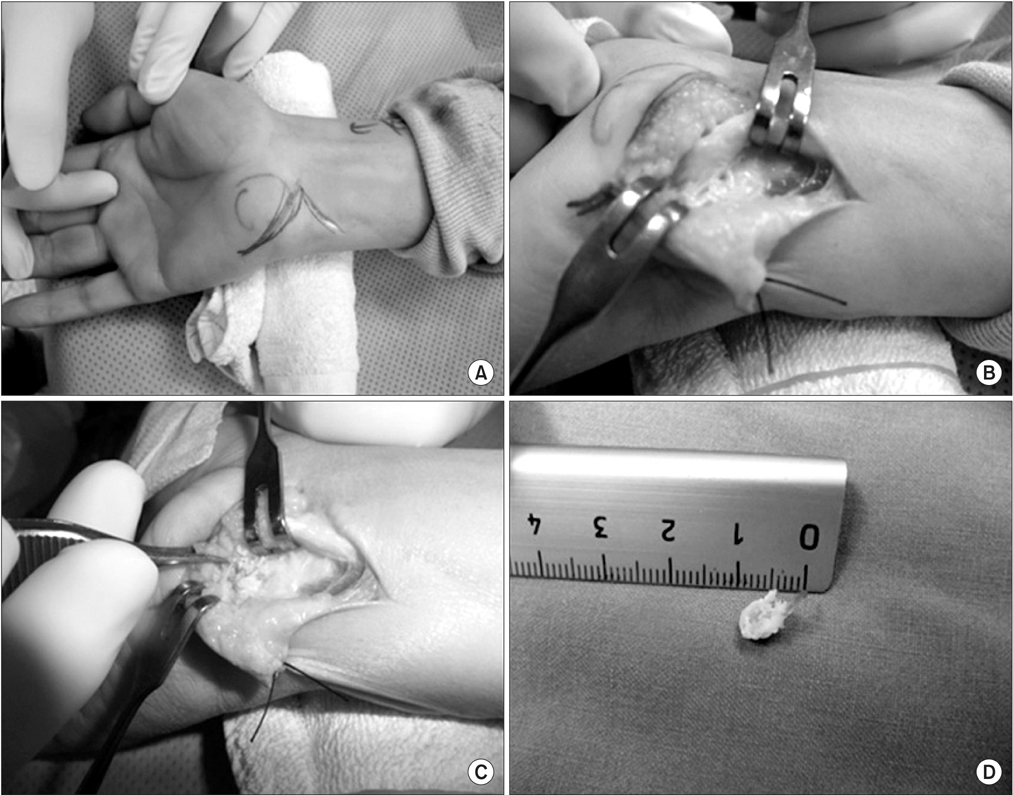
Figure 6 If symptoms persist despite aggressive nonsurgical management, operative treatment may be indicated. (A) The flexor carpi ulnaris is usually approached through a zigzag incision directly over the flexor carpi ulnaris, extending distally over the pisiform. (B) The flexor carpi ulnaris is identified and retracted radially. (C) The crystalline material and any degenerative tendon and inflammatory tissue are then debrided. (D) Resected crystallin material and hypertrophied pisiform are shown (right lower).
Reference
-
1. Sachar K. Ulnar-sided wrist pain: evaluation and treatment of triangular fibrocartilage complex tears, ulnocarpal impaction syndrome, and lunotriquetral ligament tears. J Hand Surg Am. 2012; 37:1489–1500.2. Taleisnik J, Gelberman RH, Miller BW, Szabo RM. The extensor retinaculum of the wrist. J Hand Surg Am. 1984; 9:495–501.3. Spinner M, Kaplan EB. Extensor carpi ulnaris. Its relationship to the stability of the distal radio-ulnar joint. Clin Orthop Relat Res. 1970; 68:124–129.4. Allende C, Le Viet D. Extensor carpi ulnaris problems at the wrist: classification, surgical treatment and results. J Hand Surg Br. 2005; 30:265–272.5. Inoue G, Tamura Y. Surgical treatment for recurrent dislocation of the extensor carpi ulnaris tendon. J Hand Surg Br. 2001; 26:556–559.6. Oka Y, Handa A. Recurrent dislocation of the ECU tendon in a golf player: release of the extensor retinaculum and partial resection of the ulno-dorsal ridge of the ulnar head. Hand Surg. 2001; 6:227–230.7. MacLennan AJ, Nemechek NM, Waitayawinyu T, Trumble TE. Diagnosis and anatomic reconstruction of extensor carpi ulnaris subluxation. J Hand Surg Am. 2008; 33:59–64.8. Nakashima T, Hojo T, Furukawa H. Deep and shallow forms of the sulcus for extensor carpi ulnaris. J Anat. 1993; 183:635–638.9. Melone CP Jr, Nathan R. Traumatic disruption of the triangular fibrocartilage complex. Pathoanatomy. Clin Orthop Relat Res. 1992; (275):65–73.10. Montalvan B, Parier J, Brasseur JL, Le Viet D, Drape JL. Extensor carpi ulnaris injuries in tennis players: a study of 28 cases. Br J Sports Med. 2006; 40:424–429. discussion 429.11. Jeantroux J, Becce F, Guerini H, Montalvan B, Le Viet D, Drapé JL. Athletic injuries of the extensor carpi ulnaris subsheath: MRI findings and utility of gadolinium-enhanced fat-saturated T1-weighted sequences with wrist pronation and supination. Eur Radiol. 2011; 21:160–166.12. Wang C, Gill TJ 4th, Zarins B, Herndon JH. Extensor carpi ulnaris tendon rupture in an ice hockey player: a case report. Am J Sports Med. 2003; 31:459–461.13. Vulpius J. Habitual dislocation of the extensor carpri ulnaris tendon. Acta Orthop Scand. 1964; 34:105–108.14. Rayan GM. Recurrent dislocation of the extensor carpi ulnaris in athletes. Am J Sports Med. 1983; 11:183–184.15. Hajj AA, Wood MB. Stenosing tenosynovitis of the extensor carpi ulnaris. J Hand Surg Am. 1986; 11:519–520.16. Dickson DD, Luckey CA. Tenosynovitis of the extensor carpi ulnaris tendon sheath. J Bone Joint Surg Am. 1948; 30A:903–907.17. Kip PC, Peimer CA. Release of the sixth dorsal compartment. J Hand Surg Am. 1994; 19:599–601.18. Nachinolcar UG, Khanolkar KB. Stenosing tenovaginitis of extensor carpi ulnaris: brief report. J Bone Joint Surg Br. 1988; 70:842.19. Dilley DF, Tonkin MA. Acute calcific tendinitis in the hand and wrist. J Hand Surg Br. 1991; 16:215–216.20. Budoff JE, Kraushaar BS, Ayala G. Flexor carpi ulnaris tendinopathy. J Hand Surg Am. 2005; 30:125–129.21. Wick MC, Weiss RJ, Arora R, et al. Enthesiopathy of the flexor carpi ulnaris at the pisiform: findings of high-frequency sonography. Eur J Radiol. 2011; 77:240–244.22. Kwon SM, Cha JH, Oh JR. Surgical management of pisiform bone deformity associated with tendonitis of flexor carpi ulnaris. J Korean Soc Surg Hand. 2013; 18:132–137.23. Palmieri TJ. Pisiform area pain treatment by pisiform excision. J Hand Surg Am. 1982; 7:477–480.24. Lam KS, Woodbridge S, Burke FD. Wrist function after excision of the pisiform. J Hand Surg Br. 2003; 28:69–72.25. Carroll RE, Coyle MP Jr. Dysfunction of the pisotriquetral joint: treatment by excision of the pisiform. J Hand Surg Am. 1985; 10:703–707.26. Gómez CL, Renart IP, Pujals JI, Palou EC, Busquets RC. Dysfunction of the pisotriquetral joint: degenerative arthritis treated by excision of the pisiform. Orthopedics. 2005; 28:405–408.
- Full Text Links
-
- Actions
-
Cited
- CITED
-
- Close
- Share
-
- Similar articles
-
- Anatomical Relation of Ulnar Nerve and Flexor Carpi Ulnaris Muscle at the Wrist
- Clinical Study of Kienbock's Disease
- Ulnar Neuropathy Caused by a Schwannoma in the Guyon's Cannal
- Ulnar Nerve Compression at Guyon's Canal by an Arteriovenous Malformation
- Ulnar nerve Compression Syndrome due to anomalous Branch of the Ulnar Nerve Piercing the Flexor Carpi Ulnaris: Report of one case
