A Case of Sclerosed Hemangioma Mimicking Intrahepatic Cholangiocarcinoma
- Affiliations
-
- 1Department of Gastroenterology, Fatima Hospital, Daegu, Korea. murmpyo@yahoo.co.kr
- 2Department of General Surgery, Fatima Hospital, Daegu, Korea.
- 3Department of Radiology, Fatima Hospital, Daegu, Korea.
- 4Department of Pathology, Fatima Hospital, Daegu, Korea.
- KMID: 1775941
- DOI: http://doi.org/10.4166/kjg.2009.54.6.399
Abstract
- Hemangioma is one of the most frequently encountered benign hepatic neoplasm which can develop secondary degeneration. Sclerosed hemangioma is a rare disease histologically characterized by large amount of collagen and elastic fibril between sclerosed small vessels. Its differential diagnosis is very difficult. It should be included in the differential diagnosis of other hepatic lesions such as hepatocellular carcinoma, intrahepatic cholangiocarcinoma, and metastatic hepatic tumor. A 77-year old male was admitted with upper abdominal discomfort. Abdominal ultrasonography revealed GB stone, dilated common bile duct with bile duct stone, and a 4.6 cm sized hyperechoic mass at segment 5 and 6 of the liver. Abdominal dynamic computed tomography demonstrated dilated intrahepatic bile ducts and a 5x5 cm sized mass which showed minimally delayed enhancement. Abdominal magnetic resonance imaging revealed the mass with low signal intensity in T1 weighted image, high signal intensity and focal low signal in T2 weighted image which showed minimal enhancement. We removed common bile duct stone with endoscopic retrograde cholangiopancreatography then decided to undergo right lower segmentectomy of liver due to possibility of cholangiocarcinoma. Histopathological examination of hepatic mass showed large amount of fibrous tissue with occasional residual vascular channels. We describe one case of sclerosed hemangioma mimicking cholangiocarcinoma.
Keyword
MeSH Terms
Figure
-
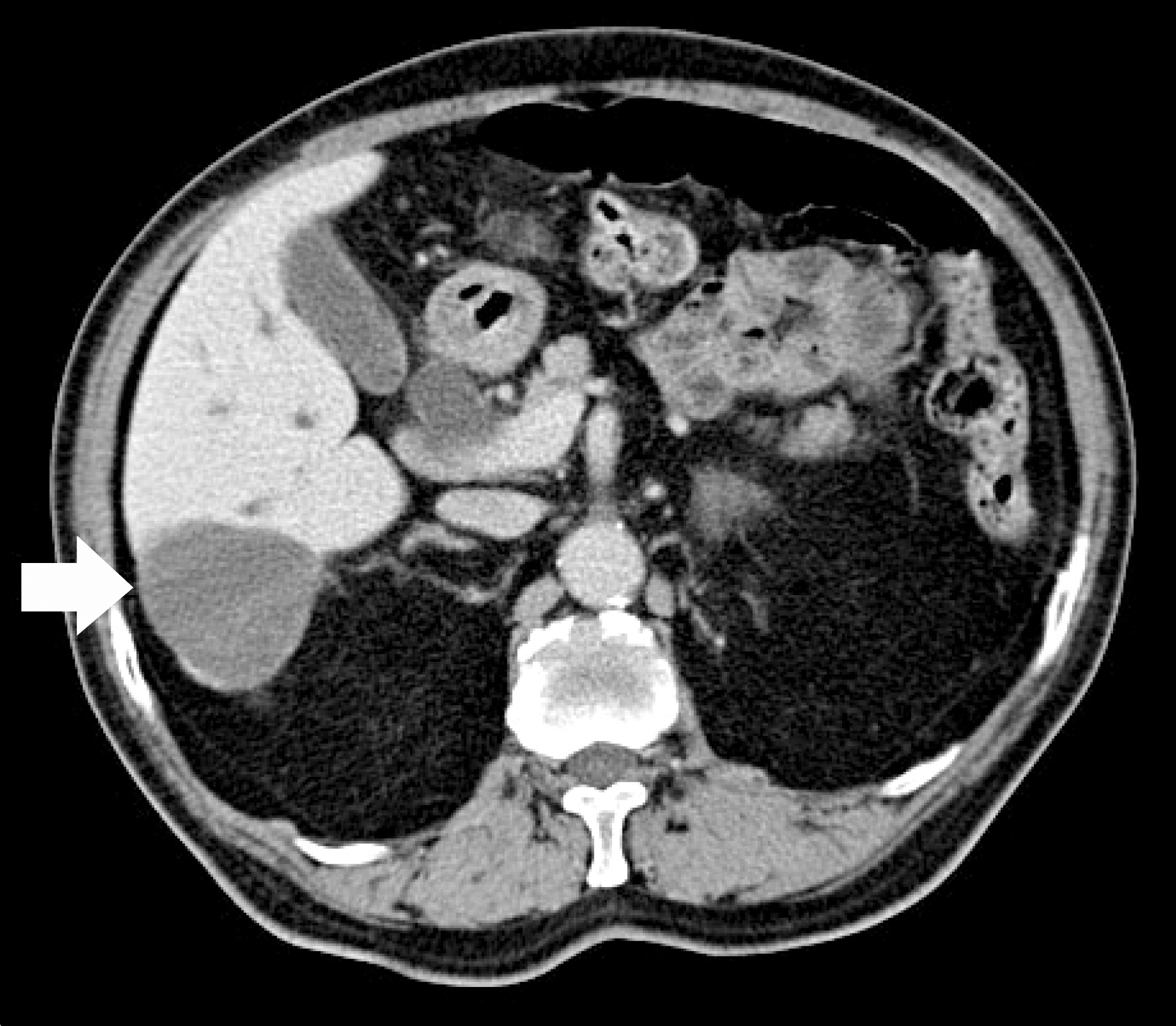
Fig. 1. Abdominal computed tomography scan revealed large, elliptical, and well marginated mass in the right lower lobe of the liver with delayed partial enhancement.

Fig. 2. Magnetic resonance imaging. (A) T1-weighted image showed a large and moderately hypointense mass. (B) T2-weighted image image showed a large, and predominant hyperintense and partial hypointense mass.
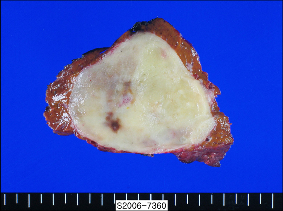
Fig. 3. Macroscopic view of the resected tumor showed a well demarcated pale yellow to grayish soft mass with focal brownish hemorrhage.
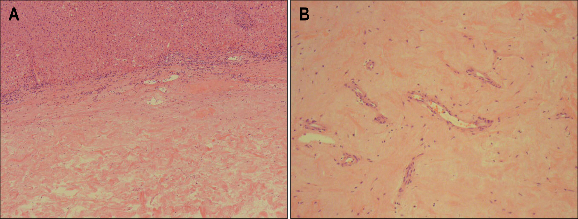
Fig. 4. Microscopic view of the resected tumor. (A) Low magnification showed a large amount of fibrous tissue with occasional residual vascular channels (H&E stain, ×20). (B) Higher magnification showed dense fibrous tissue between small vascular channels lined with endothelial cells (H&E stain, ×100).
Reference
-
1. Ishak KG, Rabin L. Benign tumors of the liver. Med Clin North Am. 1975; 59:995–1013.
Article2. Stark DD, Felder RC, Wittenberg J, et al. Magnetic resonance imaging of cavernous hemangioma of the liver: tissue-specific characterization. Am J Roentgenol. 1985; 145:213–222.
Article3. Nelson RC, Chezmar JL. Diagnostic approach to hepatic hemangiomas. Radiology. 1990; 176:11–13.
Article4. Semelka RC, Brown ED, Ascher SM, et al. Hepatic hemangiomas: a multi-institutional study of appearances on T2-weighted and serial gadolinium-enhanced gradient-echo MR images. Radiology. 1994; 192:401–406.5. Choi BI, Han MC, Kim CW. Small hepatocellular carcinoma versus small cavernous hemangioma: differentiation with MR imaging at 2.0 T. Radiology. 1990; 176:103–106.
Article6. Goldman ZD. Benign tumors of the liver. Okuda K, Ishak KG, editors. eds.Neoplasm of the liver. Volume 1. 1st ed.Tokyo: Springer-Verlag;1987. p. 105–125.7. Vilgrain V, Boulos L, Vullierme MP, Denys A, Terris B, Menu Y. Imaging of atypical hemangiomas of the liver with pathologic correlation. Radiographics. 2000; 20:379–397.
Article8. Cheng HC, Tsai SH, Chiang JH, Chang CY. Hyalinized liver hemangioma mimicking malignant tumor at MR imaging (letter). Am J Roentgenol. 1995; 165:1016–1017.9. Mathieu D, Rahmouni A, Vasile N, et al. Sclerosed liver hemangioma mimicking malignant tumor at MR imaging: pathologic correlation. J Magn Reason Imaging. 1994; 4:506–508.
Article10. Haratake J, Horie A, Nagafuchi Y. Hyalinized hemangioma of the liver. Am J Gastroenterol. 1992; 87:234–236.11. Takahashi K, Mulliken JB, Kozakewich HPW, Rogers RA, Folkman J, Ezekowitz AB. Cellular makers that distinguish the phases of hemangioma during infancy and childhood. J Clin Invest. 1994; 93:2357–2364.12. Yamashita Y, Shimada M, Taguchi K, et al. Hepatic sclerosing hemangioma mimicking a metastatic liver tumor. Surg Today. 2000; 30:849–852.13. Shim KS, Suh JM, Yang YS, et al. Sclerosis of hepatic cavernous hemangioma: CT findings and pathologic correlation. J Korean Med Sci. 1995; 10:294–297.
Article14. Takayasu K, Moriyama N, Shima Y, et al. Atypical radiographic findings in hepatic cavernous hemangioma: correlation with histologic features. AJR Am J Roentgenol. 1986; 146:1149–1153.
Article15. Doyle DJ, Khalili K, Guindi M, Atri M. Imaging features of sclerosed hemangioma. AJR Am J Roentgenol. 2007; 189:67–72.
Article16. Hiroki M, Toru I, Satoru I, et al. Sclerosed hemangioma of the liver: report of a case and review of the literature. Hepatology Research. 2008; 38:529–533.
Article
- Full Text Links
-
- Actions
-
Cited
- CITED
-
- Close
- Share
-
- Similar articles
-
- Sclerosed Hemangioma of the Liver
- Sclerosed hemangioma of the liver
- Primary hepatic tuberculosis mimicking intrahepatic cholangiocarcinoma: report of two cases
- A Case of Dermatomyositis Associated with Infiltrative Intrahepatic Cholangiocarcinoma
- A Case of Intrahepatic Cholangiocarcinoma Developed in a Remote Region from the Site of Hepatolithiasis
