Phase Contrast Microradiography of Mouse Lung Using Synchrotron X-ray: Correlation with Optical Microscopy
- Affiliations
-
- 1Department of Radiology, Korea University Guro Hospital, Seoul, Korea. keyrad@korea.ac.kr
- 2Department of Pathology, Korea University Guro Hospital, Seoul, Korea.
- 3The Research Institute for Skin Imaging, Korea University Guro Hospital, Seoul, Korea.
- 4Department of Material Science and Engineering, Pohang University of Science and Technology, Pohang, Korea.
- 5Department of Radiology, Kangwon National University College of Medicine, Chuncheon, Korea.
- 6Department of Radiology, Chung-Ang University College of Medicine, Seoul, Korea.
- KMID: 1758572
- DOI: http://doi.org/10.3349/ymj.2009.50.3.422
Abstract
-
PURPOSE: The purpose of this study is to evaluate the feasibility of phase contrast X-ray microtomography and microradiography, using a polychromatic synchrotron X-ray, for analysis of the mouse lung microstructure.
MATERIALS AND METHODS
Normal mice were used for experiments. Some of the mouse lungs were prepared by the lung fixation-inflation method. The resulting sponge-like inflated lung samples were used for microtomography. The remaining mouse lungs were cut into 10 um sections and were used for microradiography and optical microscopic correlation. The experiments on mouse lung samples were performed at the 7B2 beamline of the Pohang Light Source in Korea.
RESULTS
Phase contrast X-ray microtomography of inflated lung samples showed individual alveolar structure on 3-D reconstruction. Phase contrast microradiographs of thin lung samples showed microstructure of lung, such as alveoli and bronchioles, and were well correlated with optical microscopic images.
CONCLUSIONS
The results indicate that the phase contrast X-ray microtomography and microradiography using polychromatic synchrotron X-ray is feasible for evaluation of microstructure of the lung.
Keyword
MeSH Terms
Figure
-

Fig. 1 Overall scheme of the 7B2 beamline at Pohang Light Source (PLS) and (inset) of the radiography system. The key feature of the beamline is the use of polychromatic synchrotron X-ray to achieve high lateral and time resolution by increase of X-ray flux.
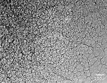
Fig. 2 Snapshot phase contrast X-ray image of inflated mouse lung sample. Overlapped alveoli are seen as an overlap pattern (left upper portion of the figure). Individual alveolar structures are clearly defined at the peripheral portion of tissue sample where overlapping of alveoli is minimized (right lower portion of the figure).
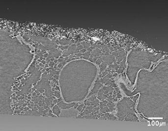
Fig. 3 On 3-D reconstruction of phase contrast X-ray microtomography, the individual alveolar structures and airways of mouse lung were clearly defined.

Fig. 4 Phase contrast microradiograph (A) of 10 µm thickness section of mouse lung shows very fine microstructure of respiratory epitheliums (small arrows) and bronchial cartilage (large arrow). This microradiograph correlates well with optical microscopic image (B).

Fig. 5 Phase contrast microradiograph (A) of 10 µm thickness section of mouse lung shows very fine microstructure of alveoli (asterisk), bronchiole (small arrow), and small muscular arteries (large arrow). This microradiograph correlates well with microscopic image (B).
Reference
-
1. Gao D, Pogany A, Stevenson AW, Wilkins SW. Phase-contrast radiography. Radiographics. 1998. 18:1257–1267.2. Meuli R, Hwu Y, Je JH, Margaritondo G. Synchrotron radiation in radiology: radiology techniques based on synchrotron sources. Eur Radiol. 2004. 14:1550–1560.3. Lewis RA. Medical phase contrast x-ray imaging: current status and future prospects. Phys Med Biol. 2004. 49:3573–3583.
Article4. Kitchen MJ, Paganin D, Lewis RA, Yagi N, Uesugi K, Mudie ST. On the origin of speckle in x-ray phase contrast images of lung tissue. Phys Med Biol. 2004. 49:4335–4348.
Article5. Kitchen MJ, Lewis RA, Yagi N, Uesugi K, Paganin D, Hooper SB, et al. Phase contrast X-ray imaging of mice and rabbit lungs: a comparative study. Br J Radiol. 2005. 78:1018–1027.
Article6. Lewis RA, Yagi N, Kitchen MJ, Morgan MJ, Paganin D, Siu KK, et al. Dynamic imaging of the lungs using x-ray phase contrast. Phys Med Biol. 2005. 50:5031–5040.
Article7. Jheon S, Youn HS, Kim HT, Choi GH, Kim JK. High-resolution X-ray refraction imaging of rat lung and histological correlations. Microsc Res Tech. 2006. 69:656–659.8. Sera T, Uesugi K, Yagi N. Refraction-enhanced tomography of mouse and rabbit lungs. Med Phys. 2005. 32:2787–2792.
Article9. Start A. Groskint. Heintzman's The Lung: Radiological-Pathological Correlations. 1993. St.Louis: Mosby.10. Hwu Y, Tsai WL, Je JH, Seol SK, Kim B, Groso A, et al. Synchrotron microangiography with no contrast agent. Phys Med Biol. 2004. 49:501–508.
Article11. Tong Y, Zhang G, Li Y, Tan M, Wang W, Chen J, et al. Synchrotron microradiography study on acute lung injury of mouse caused by PM(2.5) aerosols. Eur J Radiol. 2006. 58:266–272.12. Watz H, Breithecker A, Rau WS, Kriete A. Micro-CT of the human lung: imaging of alveoli and virtual endoscopy of an alveolar duct in a normal lung and in a lung with centrilobular emphysema--initial observations. Radiology. 2005. 236:1053–1058.13. Langheinrich AC, Leithäuser B, Greschus S, Von Gerlach S, Breithecker A, Matthias FR, et al. Acute rat lung injury: feasibility of assessment with micro-CT. Radiology. 2004. 233:165–171.
- Full Text Links
-
- Actions
-
Cited
- CITED
-
- Close
- Share
-
- Similar articles
-
- Synchrotron Radiation Imaging of Breast Tissue Using a Phase-contrast Hard X-ray Microscope
- Evaluation of Phase-Contrast Microscopic Imaging with Synchrotron Radiation in the Diagnosis of Breast Cancer and Differentiation of Various Breast Diseases: Preliminary Results
- Synchrotron Radiation Imaging of Female Breast Tissues Using Phase Contrast Technique
- 3D Histology Using the Synchrotron Radiation Propagation Phase Contrast Cryo-microCT
- Assessment of Early Dental Caries by Using Optical Coherence Tomography
