A New Method of Measuring the Amount of Soft Tissue in Pulmonary Ground-Glass Opacity Nodules: a Phantom Study
- Affiliations
-
- 1Department of Radiology, Seoul National University Bundang Hospital, Gyeonggi-go, Korea. lkwrad@radiol.snu.ac.kr
- 2Department of Radiology, Seoul National University Hospital, Seoul, Korea.
- KMID: 1758455
- DOI: http://doi.org/10.3348/kjr.2008.9.3.219
Abstract
OBJECTIVE
To devise a new method to measure the amount of soft tissue in pulmonary ground-glass opacity nodules, and to compare the use of this method with a previous volumetric measurement method by use of a phantom study. MATERIALS AND METHODS: Phantom nodules were prepared with material from fixed normal swine lung. Forty nodules, each with a diameter of 10 mm, were made with a variable mean attenuation. The reference-standard amount of soft tissue in the nodules was obtained by dividing the weight by the specific gravity. The imaging data on the phantom nodules were acquired with the use of a 16-channel multidetector CT scanner. The CT-measured amount of soft tissue of the nodules was calculated as follows: soft tissue amount = volume x (1 + mean attenuation value / 1,000). The relative percentage error (RPE) between the CT-measured amount of the soft tissue and the reference-standard amount of the soft tissue was also measured. The RPEs determined with use of the new method were compared with the RPEs determined with the current volumetric measurement method by the use of the paired t test. RESULTS: The CT-measured amount of soft tissue showed a strong correlation with the reference-standard amount of soft tissue (R(2) = 0.996, p < 0.01). The mean RPE of the CT-measured amount of soft tissue in the nodules was -7.79 +/- 1.88%. The mean RPE of the CT-measured volume was 114.78 +/- 51.02%, which was significantly greater than the RPE of the CT-measured amount of soft tissue (p < 0.01). CONCLUSION: The amount of soft tissue measured by the use of CT reflects the reference-standard amount of soft tissue in the ground-glass opacity nodules much more accurately than does the use of the CT-measured volume.
MeSH Terms
Figure
-
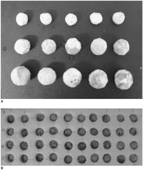
Fig. 1 Phantom nodules made of fixed swine lung (A). Forty nodules were placed in wells with diameter of approximately 10 mm of 5 cm-thick Styrofoam plate (B).
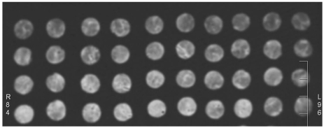
Fig. 2 CT image of 40 ground-glass opacity nodules. Mean attenuation values of nodules range from -754 HU to -352 HU.
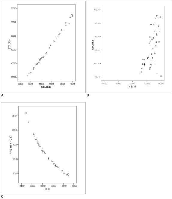
Fig. 3 Scatterplots of linear regression.A. Regression of STARS on STACT of nodules at -900 HU lower threshold level. Scatterplot shows very strong correlation between STACT and STARS. Resulting linear equation was as follows: STARS = 1.036 × STACT + 21.199 (R2 = 0.996, p < 0.01).B. Regression of STARS on VCT of nodules at -900 HU lower threshold level. Scatterplot shows poor correlation between VCT and STARS (R2 = 0.294, p < 0.01).C. Regression of relative percent error of VCT on attenuation values of nodules at 900 HU lower threshold level. Scatterplot shows negative strong correlation between mean attenuation value of nodules and VCT (R2 = 0.941, p < 0.01).RPE = relative percentage error; STACT = CT-measured amount of soft tissue in nodules; STARS = reference-standard amount of soft tissue in nodules; VCT = CT-measured volume of nodules; MHU = mean attenuation value of nodules
Reference
-
1. Diederich S, Wormanns D, Semik M, Thomas M, Lenzen H, Roos N, et al. Screening for early lung cancer with low-dose spiral CT: prevalence in 817 asymptomatic smokers. Radiology. 2002; 222:773–781. PMID: 11867800.
Article2. Henschke CI, Yankelevitz DF, Mirtcheva R, McGuinness G, McCauley D, Miettinen OS. CT screening for lung cancer: frequency and significance of part-solid and nonsolid nodules. AJR Am J Roentgenol. 2002; 178:1053–1057. PMID: 11959700.3. Yankelevitz DF, Reeves AP, Kostis WJ, Zhao B, Henschke CI. Small pulmonary nodules: volumetrically determined growth rates based on CT evaluation. Radiology. 2000; 217:251–256. PMID: 11012453.
Article4. Wormanns D, Kohl G, Klotz E, Marheine A, Beyer F, Heindel W, et al. Volumetric measurements of pulmonary nodules at multi-row detector CT: in vivo reproducibility. Eur Radiol. 2004; 14:86–92. PMID: 14615902.
Article5. Revel M-P, Lefort C, Bissery A, Bienvenu M, Aycard L, Chatellier G, et al. Pulmonary nodules: preliminary experience with three-dimensional evaluation. Radiology. 2004; 231:459–466. PMID: 15128991.
Article6. Goo JM, Lee JW, Lee HJ, Kim S, Kim JH, Im JG. Automatic lung nodule detection at low-dose CT: preliminary experience. Korean J Radiol. 2003; 4:211–216. PMID: 14726637.7. Aoki T, Nakata H, Watanabe H, Nakamura K, Kasai T, Hashimoto H, et al. Evolution of peripheral lung adenocarcinomas: CT findings correlated with histology and tumor doubling time. AJR Am J Roentgenol. 2000; 174:763–768. PMID: 10701622.8. Takashima S, Maruyama Y, Hasegawa M, Yamanda T, Honda T, Kadoya M, et al. CT findings and progression of small peripheral lung neoplasms having a replacement growth pattern. AJR Am J Roentgenol. 2003; 180:817–826. PMID: 12591704.
Article9. Kim EA, Johkoh T, Lee KS, Han J, Fujimoto K, Sadohara J, et al. Quantification of ground-glass opacity on high-resolution CT of small peripheral adenocarcinoma of the lung: pathologic and prognostic implications. AJR Am J Roentgenol. 2001; 177:1417–1422. PMID: 11717098.10. Lee HJ, Goo JM, Lee CH, Yoo CG, Kim YT, Im JG. Nodular ground-glass opacities on thin-section CT: size change during follow-up and pathological results. Korean J Radiol. 2007; 8:22–31. PMID: 17277560.
Article11. Ko JP, Rusinek H, Jacobs EL, Babb JS, Betke M, McGuinness G, et al. Small pulmonary nodules; volume measurement at chest CT-phantom study. Radiology. 2003; 228:864–870. PMID: 12954901.
Article12. Markarian B, Dailey ET. Heizman ER, editor. Preparation of inflated lung specimens. The Lung : radiologic-pathologic correlations. 1984. 2nd ed. St Louis, MO: Mosby-Year Book;p. 4–12.13. Vieira SR, Puybasset L, Lu Q, Richecoeur J, Cluzel P, Coriat P, et al. A scanographic assessment of pulmonary morphology in acute lung injury. Significance of the lower inflection point detected on the lung pressure-volume curve. Am J Respir Crit Care Med. 1999; 159:1612–1623. PMID: 10228135.14. Lee HJ, Im JG, Goo JM, Kim YI, Lee MW, Ryu HG, et al. Acute lung injury: effects of prone positioning on cephalocaudal distribution of lung inflation CT assessment in dogs. Radiology. 2005; 234:151–161. PMID: 15550370.15. Malbouisson LM, Muller JC, Constantin JM, Lu Q, Puybasset L, Rouby JJ. CT Scan ARDS Study Group. Computed tomography assessment of positive end-expiratory pressure-induced alveolar recruitment in patients with acute respiratory distress syndrome. Am J Respir Crit Care Med. 2001; 163:1444–1450. PMID: 11371416.16. Marten K, Funke M, Engelke C. Flat panel detector-based volumetric CT: prototype evaluation with volumetry of small artificial nodules in a pulmonary phantom. J Thorac Imaging. 2004; 19:156–163. PMID: 15273611.17. Do KH, Chung MJ, Goo JM, Lee KW, Im JG. Evaluation of computer aided volumetry for simulated small pulmonary nodules on computed tomography. J Korean Radiol Soc. 2004; 50:101–108.
Article18. Henschke CI, Naidich DP, Yankelevitz DF, McGuinness G, McCauley DI, Smith JP, et al. Early lung cancer action project: initial findings on repeat screening. Cancer. 2001; 92:153–159. PMID: 11443621.19. Jennings SG, Winer-Muram HT, Traver RD, Farber MO. Lung tumor growth: assessment with CT comparison of diameter and cross-sectional area with volume measurement. Radiology. 2004; 231:866–871. PMID: 15163822.
- Full Text Links
-
- Actions
-
Cited
- CITED
-
- Close
- Share
-
- Similar articles
-
- The Clinical Approach to Nodular Ground Glass Opacity in the Lung
- Radiographic Findings of Miliary Tuberculosis: Difference in Patients with and those without Associated Acute Respiratory Failure
- Radiologic Approach to the Idiopathic Interstitial Pneumonias
- Paraquat Poisoning of the Lung: HRCT Findings According to the Amount of Ingestion
- Ground-Glass Opacity in Lung Metastasis from Adenocarcinoma of the Stomach: A Case Report
