MR Imaging of the Internal Auditory Canal and Inner Ear at 3T: Comparison between 3D Driven Equilibrium and 3D Balanced Fast Field Echo Sequences
- Affiliations
-
- 1Department of Radiology and Center for Imaging Science, Samsung Medical Center, Sungkyunkwan University School of Medicine, Seoul, Korea. hyungkim@skku.edu
- 2Department of Radiology, Chung-Ang University Yongsan Hospital, Seoul, Korea.
- 3Department of Radiology, Kangwon National University College of Medicine, Chuncheon, Korea.
- KMID: 1758454
- DOI: http://doi.org/10.3348/kjr.2008.9.3.212
Abstract
OBJECTIVE
To compare the use of 3D driven equilibrium (DRIVE) imaging with 3D balanced fast field echo (bFFE) imaging in the assessment of the anatomic structures of the internal auditory canal (IAC) and inner ear at 3 Tesla (T). MATERIALS AND METHODS: Thirty ears of 15 subjects (7 men and 8 women; age range, 22-71 years; average age, 50 years) without evidence of ear problems were examined on a whole-body 3T MR scanner with both 3D DRIVE and 3D bFFE sequences by using an 8-channel sensitivity encoding (SENSE) head coil. Two neuroradiologists reviewed both MR images with particular attention to the visibility of the anatomic structures, including four branches of the cranial nerves within the IAC, anatomic structures of the cochlea, vestibule, and three semicircular canals. RESULTS: Although both techniques provided images of relatively good quality, the 3D DRIVE sequence was somewhat superior to the 3D bFFE sequence. The discrepancies were more prominent for the basal turn of the cochlea, vestibule, and all semicircular canals, and were thought to be attributed to the presence of greater magnetic susceptibility artifacts inherent to gradient-echo techniques such as bFFE. CONCLUSION: Because of higher image quality and less susceptibility artifacts, we highly recommend the employment of 3D DRIVE imaging as the MR imaging choice for the IAC and inner ear.
Keyword
MeSH Terms
Figure
-
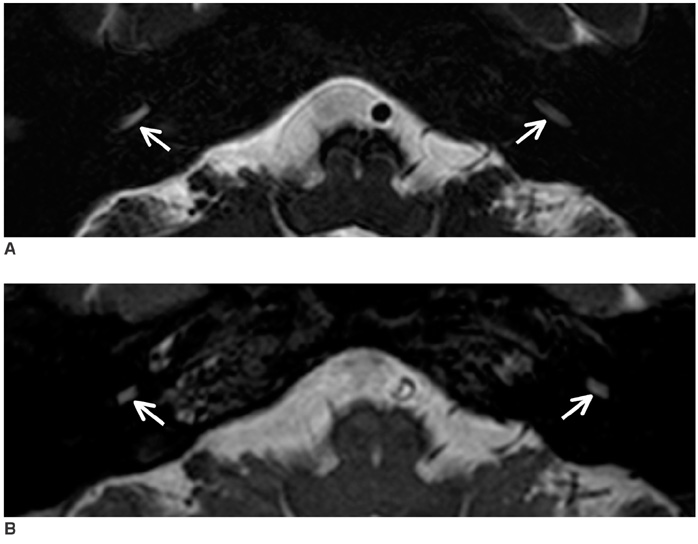
Fig. 1 Comparison of 3D driven equilibriumand 3D balanced fast field echo sequences for visualization of basal turn of cochlea. Diameter of basal turn of cochlea on both sides (arrows) is greater on 3D driven equilibrium image (A) than on 3D balanced fast field echo image (B).
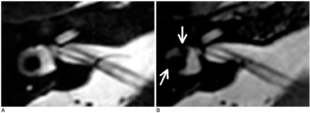
Fig. 2 Comparison of 3D driven equilibrium and 3D balanced fast field echo sequences for visualization of vestibule and lateral semicircular canal. While vestibule and lateral semicircular canal are clearly seen without any areas of signal loss on 3D driven equilibrium image (A), there are significant blackouts (arrows) due to susceptibility artifacts as seen on 3D balanced fast field echo image (B).
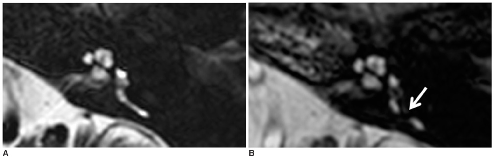
Fig. 3 Comparison of 3D driven equilibrium and 3D balanced fast field echo sequences for visualization of posterior semicircular canal. While entire posterior semicircular canal is well seen on 3D driven equilibrium image (A), there is large area of signal loss (arrow) on 3D balanced fast field echo image (B).
Reference
-
1. Casselman JW, Kuhweide R, Deimling M, Ampe W, Dehaene I, Meeus L. Constructive interference in steady state-3DFT MR imaging of the inner ear and cerebellopontine angle. AJNR Am J Neuroradiol. 1993. 14:47–57.2. Held P, Fellner C, Fellner F, Seitz L, Graf S, Hilbert M, et al. MRI of inner ear and facial nerve pathology using 3D MPRAGE and 3D CISS sequences. Br J Radiol. 1997. 70:558–566.3. Naganawa S, Ito T, Fukatsu H, Ishigaki T, Nakashima T, Ichinose N, et al. MR imaging of the inner ear: comparison of a three-dimensional fast spin-echo sequence with use of a dedicated quadrature-surface coil with a gadolinium-enhanced spoiled gradient-recalled sequence. Radiology. 1998. 208:679–685.4. Yang D, Kodama T, Tamura S, Watanabe K. Evaluation of the inner ear by 3D fast asymmetric spin echo (FASE) MR imaging: phantom and volunteer studies. Magn Reson Imaging. 1999. 17:171–182.5. Schmalbrock P. Comparison of three-dimensional fast spin echo and gradient echo sequences for high-resolution temporal bone imaging. J Magn Reson Imaging. 2000. 12:814–825.6. Naganawa S, Koshikawa T, Fukatsu H, Ishigaki T, Fukuta T. MR cisternography of the cerebellopontine angle: comparison of three-dimensional fast asymmetrical spin-echo and three-dimensional constructive interference in the steady-state sequences. AJNR Am J Neuroradiol. 2001. 22:1179–1185.7. Nakashima K, Morikawa M, Ishimaru H, Ochi M, Kabasawa H, Hayashi K. Three-dimensional fast recovery fast spin-echo imaging of the inner ear and the vestibulocochlear nerve. Eur Radiol. 2002. 12:2776–2780.8. Nakamura T, Naganawa S, Koshikawa T, Fukatsu H, Sakurai Y, Aoki I, et al. High-spatial-resolution MR cisternography of the cerebellopontine angle in 90 seconds with a zero-fill interpolated fast recovery 3D fast asymmetric spin-echo sequence. AJNR Am J Neuroradiol. 2002. 23:1407–1412.9. Ciftci E, Anik Y, Arslan A, Akansel G, Sarisoy T, Demirci A. Driven equilibrium (drive) MR imaging of the cranial nerves V-VIII: comparison with the T2-weighted 3D TSE sequence. Eur J Radiol. 2004. 51:234–240.10. Tsuchiya K, Aoki C, Hachiya J. Evaluation of MR cisternography of the cerebellopontine angle using a balanced fast-fieldecho sequence: preliminary findings. Eur Radiol. 2004. 14:239–242.11. Naganawa S, Koshikawa T, Fukatsu H, Ishigaki T, Aoki I, Ninomiya A. Fast recovery 3D fast spin-echo MR imaging of the inner ear at 3T. AJNR Am J Neuroradiol. 2002. 23:299–302.12. Lane JI, Ward H, Witte RJ, Bernstein MA, Driscoll CL. 3-T imaging of the cochlear nerve and labyrinth in cochlear-implant candidates: 3D fast recovery fast spin-echo versus 3D constructive interference in the state techniques. AJNR Am J Neuroradiol. 2004. 25:618–622.13. Jung NY, Moon WJ, Lee MH, Chung EC. Magnetic resonance cisternography: comparison between 3-dimensional driven equilibrium with sensitivity encoding and 3-dimensional balanced fast-field echo sequences with sensitivity encoding. J Comput Assist Tomogr. 2007. 31:588–591.14. Melhem ER, Itoh R, Folkers PJ. Cervical spine: three-dimensional fast spin-echo MR imaging-improved recovery of longitudinal magnetization with driven equilibrium pulse. Radiology. 2001. 218:283–288.15. Preibisch C, Pilatus U, Bunke J, Hoogenraad F, Zanella F, Lanfermann H. Functional MRI using sensitivity-encoded echo planar imaging (SENSE-EPI). Neuroimage. 2003. 19:412–421.16. Constable RT, Gore JC. The loss of small objects in variable TE imaging: implications for FSE, RARE, and EPI. Magn Reson Med. 1992. 28:9–24.17. Ross JS. The high-field-strength curmudgeon. AJNR Am J Neuroradiol. 2004. 25:168–169.18. Tanenbaum LN. 3-T MR imaging: ready for clinical practice. AJNR Am J Neuroradiol. 2004. 25:1626–1627.
- Full Text Links
-
- Actions
-
Cited
- CITED
-
- Close
- Share
-
- Similar articles
-
- T1-weighted MR Imaging of the Neonatal Brain at 3.0 Tesla: Comparison of Spin Echo, Fast Inversion Recovery, and Magnetization-prepared Three Dimensional Gradient Echo Techniques
- A New Promising Technique of 3D Isovoxel Imaging Using 3T MRI in the Wrist: Comparison with 3T MR Arthrography
- Single-Slab 3D Fast Spin Echo and Its Variants With Very Long Echo Trains for Clinical T 2 -Weighted Contrast
- Fast Imaging of Shoulder MR Arthrography With Compressed Sensing Accelerated Isovolumetric 3D-THRIVE Sequence: Comparing One Isovolumetric Scan With Multiplanar Reconstruction and Three Conventional MR Images
- Qualitative and Quantitative Assessment of Isotropic Ankle Magnetic Resonance Imaging: Three-Dimensional Isotropic Intermediate-Weighted Turbo Spin Echo versus Three-Dimensional Isotropic Fast Field Echo Sequences
