A New Promising Technique of 3D Isovoxel Imaging Using 3T MRI in the Wrist: Comparison with 3T MR Arthrography
- Affiliations
-
- 1Department of Radiology, Korea University Ansan Hospital, Korea University College of Medicine, Gyeonggi-do, Korea. kimbaekh@hanmail.net
- 2Department of Orthopedic Surgery, Korea University Ansan Hospital, Korea University College of Medicine, Gyeonggi-do, Korea.
- KMID: 2097936
- DOI: http://doi.org/10.3348/jksr.2011.64.2.185
Abstract
- PURPOSE
We wanted to evaluate the usefulness of 3D isovoxel MR imaging using 3T MRI in the wrist joint, as compared with 3T MR arthrography.
MATERIALS AND METHODS
A total of 33 patients underwent both MR arthrography and 3D isovoxel imaging of the wrist joints using 3T MR, including 11 patients with arthroscopic confirmation. 3D isovoxel MR imaging was performed using an intermediate-weighted fast spin echo coronal scan with a 0.4-mm slice thickness and the axial images were reconstructed with a 1-mm slice thickness. One radiologist evaluated for the presence of scapholunate or lunotriquetral ligament tear and she determined the grade of the triangular fibrocartilage complex tear and chondromalacia with its location. We compared the two examinations using kappa values.
RESULTS
The rates of detecting wrist injury were similar for both exams with substantial to almost perfect inter-examination agreement (kappa value = 0.864 for scapholunate ligament tear, 0.835 for lunotriquetral ligament tear, 0.799 for TFCC tear and 0.940 for chondromalacia). For the eleven cases that underwent arthroscopy, their results of 3D isovoxel MRI were also similar to that of MR arthrography.
CONCLUSION
3D isovoxel MR imaging is useful for the evaluation of the wrist joint.
MeSH Terms
Figure
-
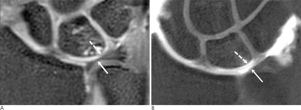
Fig. 1 A 43-year old woman with chronic wrist pain. A. A coronal 3D isovoxel MR image shows only subtle signal change in the cartilage of the lunate (dotted arrow). B. A coronal MR arthrographic image shows more clear detection of the minimal chondromalacia with marginal irregularity in the lunate (dotted arrow). Marked thinning of the triangular fibrocartilage complex with diffuse degeneration was revealed on both the 3D isovoxel MR and MR arthrography (arrows). This case was confirmed to have a grade 1 triangular fibrocartilage complex tear with grade 1 chondromalacia on arthroscopy.
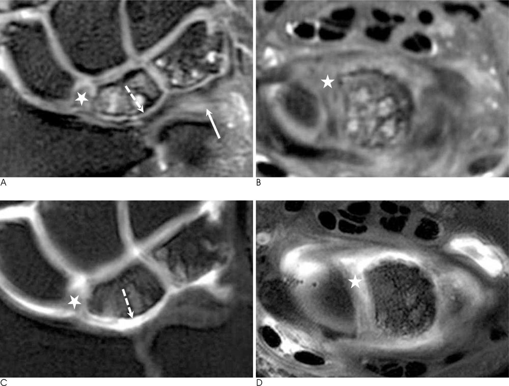
Fig. 2 A 44-year old man with chronic wrist pain. (A, C) The coronal and (B, D) axial 3D isovoxel MR and MR arthrographic images well demonstrate the scapholunte ligament tear (star). The grade 1 triangular fibrocartilage complex tear at the ulnar side (arrow) was more definite on the 3D isovoxel MR than on the MR arthrography, but grade 2 chondromalacia of the lunate (dotted arrow) was markedly seen on MR arthrography. This patient also had a lunotriquetral ligament tear seen on arthroscopy.
Reference
-
1. Khoury V, Harris PG, Cardinal E. Cross-sectional imaging of internal derangement of the wrist with arthroscopic correlation. Semin Musculoskelet Radiol. 2007; 11:36–47.2. Braun H, Kenn W, Schneider S, Graf M, Sandstede J, Hahn D. Direct MR arthrography of the wrist: value in detecting complete and partial defects of intrinsic ligaments and the TFCC in comparison with arthroscopy. Rofo. 2003; 175:1515–1524.3. Kovanlikaya I, Camili D, Cakmakci H, Goktay Y, Kovanlikaya A, Ozaksoy D, et al. Diagnostic value of MR arthrography in detection of intrinsic carpal ligament lesions: use of cine-MR arthrography as a new approach. Eur Radiol. 1997; 7:1441–1445.4. Scheck RJ, Kubitzek C, Hierner R, Szeimies U, Pfluger T, Wilhelm K, et al. The scapholunate interosseous ligament in MR arthrography of the wrist: correlation with non-enhanced MRI and wrist arthroscopy. Skeletal Radiol. 1997; 26:263–271.5. Scheck RJ, Romagnolo A, Hierner R, Pfluger T, Wilhelm K, Hahn K. The carpal ligaments in MR arthrography of the wrist: correlation with standard MRI and wrist arthroscopy. J Magn Reson Imaging. 1999; 9:468–474.6. Schmitt R, Christopoulos G, Meier R, Coblenz G, Frohner S, Lanz U, et al. Direct MR arthrography of the wrist in comparison with arthroscopy: a prospective study on 125 patients. Rofo. 2003; 175:911–919.7. Maizlin ZV, Brown JA, Clement JJ, Grebenyuk J, Fenton DM, Smith DE, et al. MR arthrogrphy of the wrist: controversies and concepts. Hand. 2009; 4:66–73.8. Palmer AK. Triangular fibrocartilage complex lesions: a classification. J Hand Surg Am. 1989; 14:594–606.9. Cerezal L, Abascal F, Garcia-Valtuille R, del Pinal F. Wrist MR arthrography: how, why, when. Radiol Clin N Am. 2005; 43:709–731.10. Tanaka T, Ogino S, Yoshioka H. Ligamentous injuries of the wrist. Semin Musculoskelet Radiol. 2008; 12:359–378.11. Joshy S, Lee K, Deshmukh SC. Accuracy of direct magnetic resonance arthrography in the diagnosis of triangular fibrocartilage complex tears of the wirst. Int Orthop. 2008; 32:251–253.12. Magee T. Can isotropic fast gradient echo imaging be substituted for conventional T1 weighted sequences in shoulder MR arthrography at 3 Tesla. J Magn Reson Imaging. 2007; 26:118–122.13. Duc SR, Pfirrmann CW, Koch PP, Zanetti M, Hodler J. Internal knee derangement assessed with 3-minute three-dimensional isovoxel true FISP MR sequence: preliminary study. Radiology. 2008; 246:526–535.14. Stevens KJ, Busse RF, Han E, Brau AC, Beatty PJ, Beaulieu CF, et al. Ankle: isotropic MR imaging with 3D-FSE-cube-initial experience in healthy volunteers. Radiology. 2008; 249:1026–1033.15. Oh DK, Yoon YC, Kwon JW, Choi SH, Jung JY, Bae S, et al. Comparison of indirect isotropic MR arthrography and conventional MR arthrography of labral lesions and rotator cuff tears: a prospective study. AJR Am J Roentgenol. 2009; 192:473–479.16. Jung JY, Yoon YC, Choi SH, Kwon JW, Yoo J, Choe BK. Three-dimensional isotropic shoulder MR arthrography: comparison with two-dimensional MR arthrography for the diagnosis fo labral lesions at 3.0 T. Radiology. 2009; 250:498–450.17. Kijowski R, Blankenbaker DG, Klaers JL, Shinki K, De Smet AA, Block WF. Vastly undersampled isotropic projection steady-state free precession imaging of the knee: diagnostic performance compared with conventional MR. Radiology. 2009; 251:185–194.
- Full Text Links
-
- Actions
-
Cited
- CITED
-
- Close
- Share
-
- Similar articles
-
- High field strength magnetic resonance imaging of musculoskeletal diseases
- High field strength magnetic resonance imaging of abdominal diseases
- Single-Dose Gadoterate Meglumine for 3T Late Gadolinium Enhancement MRI for the Assessment of Chronic Myocardial Infarction: Intra-Individual Comparison with Conventional Double-Dose 1.5T MRI
- Single-Shot Echo-Planar Diffusion-Weighted MR Imaging at 3T and 1.5T for Differentiation of Benign Vertebral Fracture Edema and Tumor Infiltration
- T1-weighted MR Imaging of the Neonatal Brain at 3.0 Tesla: Comparison of Spin Echo, Fast Inversion Recovery, and Magnetization-prepared Three Dimensional Gradient Echo Techniques
