The Quality of Reconstructed 3D Images in Multidetector-Row Helical CT: Experimental Study Involving Scan Parameters
- Affiliations
-
- 1Department of Radiology, Asan Medical Center, University of Ulsan College of Medicine, Seoul, Korea. hklee2@www.amc.seoul.kr
- 2Department of Information and Statistics,Daejeon University, Daejeon, Korea.
- KMID: 1758447
- DOI: http://doi.org/10.3348/kjr.2002.3.1.49
Abstract
OBJECTIVE
To determine which multidetector-row helical CT scanning technique provides the best-quality reconstructed 3D images, and to assess differences in image quality according to the levels of the scanning parameters used.
MATERIALS AND METHODS
Four objects with different surfaces and contours were scanned using multidetector-row helical CT at three detector-row collimations (1.25, 2.50, 5.00 mm), two pitches (3.0, 6.0), and three different degrees of overlap between the reconstructed slices (0%, 25%, 50%). Reconstructed 3D images of the resulting 72 sets of data were produced using volumetric rendering. The 72 images were graded on a scale from 1 (worst) to 5 (best) for each of four rating criteria, giving a mean score for each criterion and an overall mean score. Statistical analysis was used to assess differences in image quality according to scanning parameter levels.
RESULTS
The mean score for each rating criterion, and the overall mean score, varied significantly according to the scanning parameter levels used. With regard to detector-row collimation and pitch, all levels of scanning parameters gave rise to significant differences, while in the degree of overlap of reconstructed slices, there were significant differences between overlap of 0% and of 50% in all levels of scanning parameters, and between overlap of 25% and of 50% in overall accuracy and overall mean score. Among the 18 scanning sequences, the highest score (4.94) was achieved with 1.25 mm detector-row collimation, 3.0 pitch, and 50% overlap between reconstructed slices.
CONCLUSION
Comparison of the quality of reconstructed 3D images obtained using multidetector-row helical CT and various scanning techniques indicated that the 1.25 mm, 3.0, 50% scanning sequence was best. Quality improved as detector-row collimation decreased; as pitch was reduced from 6.0 to 3.0; and as overlap between reconstructed slices increased.
Keyword
MeSH Terms
Figure
-
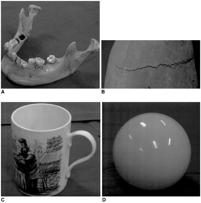
Fig. 1 The four objects used in this study. A. Mandible B. Calvarium C. Cup D. Billiard ball
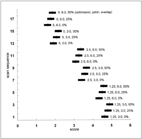
Fig. 2 Mean score and 95% confidence intervals of scanning sequences for definition.

Fig. 3 Mean score and 95% confidence intervals of scanning sequences for smoothness.

Fig. 4 Mean score and 95% confidence intervals of scanning sequences for overall accuracy.
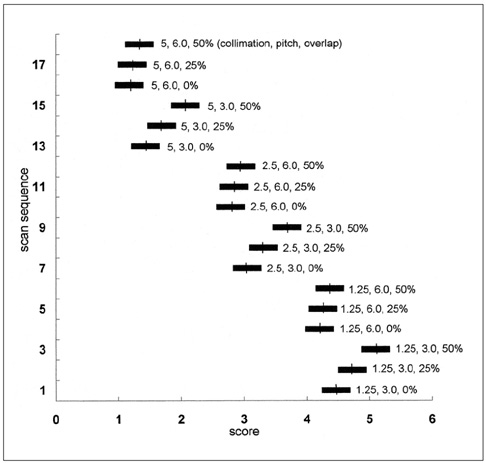
Fig. 5 Mean score and 95% confidence intervals of scanning sequences for overall mean score.

Fig. 6 Image quality of the mandible according to the level of detector-row collimation. Image quality improved as detector-row collimation decreased. A. 1.25 mm, 3.0, 50% B. 2.50 mm, 3.0, 50% C. 5.00 mm, 3.0, 50%
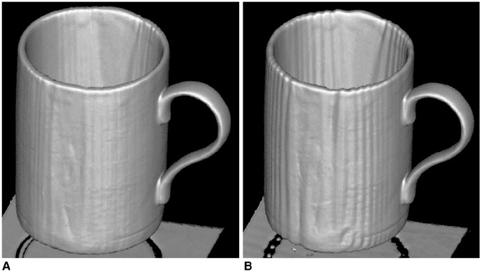
Fig. 7 Image quality of the cup according to the level of pitch. Image quality, i.e. definition of the outer margin or smoothness of the curved area, is better at a pitch of 3.0 than at one of 6.0. A. 2.50 mm, 3.0, 50% B. 2.50 mm, 6.0, 50%
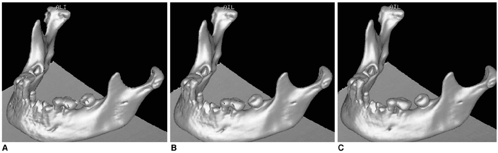
Fig. 8 Image quality of the mandible according to the degree of overlap between reconstructed slices. Image quality improved as overlap increased. A. 2.50 mm, 3.0, 0% B. 2.50 mm, 3.0, 25% C. 2.50 mm, 3.0, 50%
Reference
-
1. Kalender WA, Seissler W, Klotz E, Vock P. Spiral volumetric CT with single-breath-hold technique, continuous transport, and continuous scanner rotation. Radiology. 1990. 176:181–183.2. Crawford CR, King KF. Computed tomography scanning with simultaneous patient translation. Med Phys. 1990. 17:967–982.3. Brink JA, Heiken JP, Wang G, McEnery KW, Schlueter FJ, Vannier MW. Helical CT: principles and technical considera-tions. RadioGraphics. 1994. 14:887–893.4. Kalender WA, Polacin A. Physical performance characteristics of spiral CT scanning. Med Phys. 1991. 18:910–915.5. Liang Y, Kruger RA. Dual-slice spiral versus single-slice spiral scanning: comparison of the physical performance of two computed tomography scanners. Med Phys. 1996. 23:205–220.6. Weg N, Scheer MR, Gabor MP. Liver lesions: improved detection with dual-detector-array CT and routine 2.5-mm thin collimation. Radiology. 1998. 209:417–426.7. Hu H. Multi-slice helical CT: scan and reconstruction. Med Phys. 1999. 26:5–18.8. Taguchi K, Aradate H. Algorithm for image reconstruction in multi-slice helical CT. Med Phys. 1998. 25:550–561.9. Hu H, He HD, Foley WD, Fox SH. Multidetector-row helical CT: image quality and volume coverage speed. Radiology. 2000. 215:55–62.10. Ney DR, Fishman EK, Magid D, Robertson DD, Kawashima A. Three-dimensional volumetric display of CT data: effect of scan parameters upon image quality. J Comput Assist Tomogr. 1991. 15:875–885.11. Ney DR, Fishman EK, Kawashima A, Robertson DD Jr, Scott WW. Comparison of helical and serial CT with regard to three-dimensional imaging of musculoskeletal anatomy. Radiology. 1992. 185:865–869.12. Kasales CJ, Hopper KD, Ariola DN, et al. Reconstructed helical CT scans: improvement in z-axis resolution compared with overlapped and nonoverlapped conventional CT scans. AJR. 1995. 164:1281–1284.13. Kasales CJ, Mauger DT, Sefczek RJ, et al. Multiplanar image reconstruction and 3D imaging using a musculoskeletal phantom: conventional versus helical CT. J Comput Assist Tomogr. 1997. 21:162–169.14. Hopper KD, Pierantozzi D, Potok PS, et al. The quality of 3D reconstructions from 10 and 15 pitch helical and conventional CT. J Comput Assist Tomogr. 1996. 20:841–847.15. Kalender WA, Polacin A, Suss C. A comparison of conventional and spiral CT: an experimental study on the detection of spherical lesions. J Comput Assist Tomogr. 1994. 18:167–176.16. McEnery KW, Wilson AJ, Murphy WA Jr. Comparison of spiral computed tomography versus conventional computed tomography multiplanar reconstructions of a fracture displacement phantom. Invest Radiol. 1994. 29:665–670.17. Schoepf UJ, Bruening RD, Hong C, et al. Multislice helical CT of focal and diffuse lung disease: comprehensive diagnosis with reconstruction of contiguous and high-resolution CT sections from a single thin-collimation scan. AJR. 2001. 177:179–184.18. Lawler LP, Fishman EK. Multi-detector row CT of thoracic disease with emphasis on 3D volume rendering and CT angiography. RadioGraphics. 2001. 21:1257–1273.
- Full Text Links
-
- Actions
-
Cited
- CITED
-
- Close
- Share
-
- Similar articles
-
- Multidetector-row CT of the Gastrointestinal Tract
- Multi-Detector Row CT of the Central Airway Disease
- Traumatic Thoracic Injury: The Role of Multidetector-row CT
- Usefulness of Multidetector-row CT in the Evaluation of Reperfused Myocardial Infarction in a Rabbit Model
- Assessment of Hepatic Arterial Variation Using Multidetector Helical CT-Angiography
