Korean J Radiol.
2014 Aug;15(4):430-438. 10.3348/kjr.2014.15.4.430.
Effects of Dual-Energy CT with Non-Linear Blending on Abdominal CT Angiography
- Affiliations
-
- 1Department of Radiology, The First Affiliated Hospital of Zhengzhou University, Zhengzhou 450052, China. sulanlisll@126.com
- KMID: 1731045
- DOI: http://doi.org/10.3348/kjr.2014.15.4.430
Abstract
OBJECTIVE
To determine whether non-linear blending technique for arterial-phase dual-energy abdominal CT angiography (CTA) could improve image quality compared to the linear blending technique and conventional 120 kVp imaging.
MATERIALS AND METHODS
This study included 118 patients who had accepted dual-energy abdominal CTA in the arterial phase. They were assigned to Sn140/80 kVp protocol (protocol A, n = 40) if body mass index (BMI) < 25 or Sn140/100 kVp protocol (protocol B, n = 41) if BMI > or = 25. Non-linear blending images and linear blending images with a weighting factor of 0.5 in each protocol were generated and compared with the conventional 120 kVp images (protocol C, n = 37). The abdominal vascular enhancements, image noise, signal-to-noise ratio (SNR), contrast-to-noise ratio (CNR) and radiation dose were assessed. Statistical analysis was performed using one-way analysis of variance test, independent t test, Mann-Whitney U test, and Kruskal-Wallis test.
RESULTS
Mean vascular attenuation, CNR, SNR and subjective image quality score for the non-linear blending images in each protocol were all higher compared to the corresponding linear blending images and 120 kVp images (p values ranging from < 0.001 to 0.007) except for when compared to non-linear blending images for protocol B and 120 kVp images in CNR and SNR. No significant differences were found in image noise among the three kinds of images and the same kind of images in different protocols, but the lowest radiation dose was shown in protocol A.
CONCLUSION
Non-linear blending technique of dual-energy CT can improve the image quality of arterial-phase abdominal CTA, especially with the Sn140/80 kVp scanning.
MeSH Terms
-
Adult
Aged
Angiography/*methods
Body Mass Index
Female
Humans
Male
Middle Aged
Observer Variation
Radiation Dosage
Radiographic Image Enhancement/methods
Radiographic Image Interpretation, Computer-Assisted/*methods
Radiography, Abdominal/*methods
Signal-To-Noise Ratio
Statistics, Nonparametric
Tomography, X-Ray Computed/*methods
Figure
-
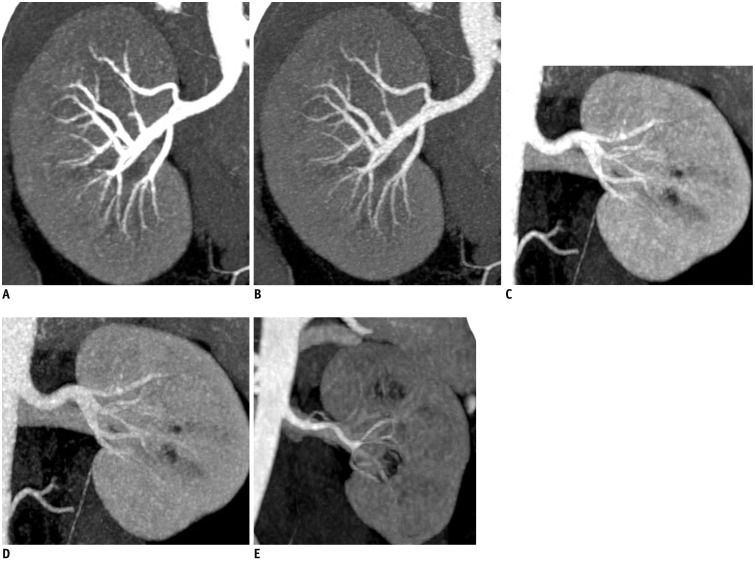
Fig. 1 Blending images and 120 kVp images. Better vascular visualization at non-linear blending images in protocol A (A) and protocol B (C) were showed compared to corresponding linear blending images [protocol A (B) and protocol B (D)] and 120 kVp images (E) (Protocol A, 80 kVp/Sn140 kVp; Protocol B, 100 kVp/Sn140 kVp).
Reference
-
1. Macovski A, Alvarez RE, Chan JL, Stonestrom JP, Zatz LM. Energy dependent reconstruction in X-ray computerized tomography. Comput Biol Med. 1976; 6:325–336. PMID: 1000958.
Article2. Alvarez RE, Macovski A. Energy-selective reconstructions in X-ray computerized tomography. Phys Med Biol. 1976; 21:733–744. PMID: 967922.3. Goo HW. CT radiation dose optimization and estimation: an update for radiologists. Korean J Radiol. 2012; 13:1–11. PMID: 22247630.
Article4. Johnson TR, Krauss B, Sedlmair M, Grasruck M, Bruder H, Morhard D, et al. Material differentiation by dual energy CT: initial experience. Eur Radiol. 2007; 17:1510–1517. PMID: 17151859.
Article5. Altenbernd J, Heusner TA, Ringelstein A, Ladd SC, Forsting M, Antoch G. Dual-energy-CT of hypervascular liver lesions in patients with HCC: investigation of image quality and sensitivity. Eur Radiol. 2011; 21:738–743. PMID: 20936520.
Article6. Marin D, Nelson RC, Samei E, Paulson EK, Ho LM, Boll DT, et al. Hypervascular liver tumors: low tube voltage, high tube current multidetector CT during late hepatic arterial phase for detection--initial clinical experience. Radiology. 2009; 251:771–779. PMID: 19346514.
Article7. Behrendt FF, Schmidt B, Plumhans C, Keil S, Woodruff SG, Ackermann D, et al. Image fusion in dual energy computed tomography: effect on contrast enhancement, signal-to-noise ratio and image quality in computed tomography angiography. Invest Radiol. 2009; 44:1–6. PMID: 19060790.8. Eusemann C, Holmes DR III, Schmidt B, Flohr TG, Robb R, McCollough C, et al. Dual energy CT: how to best blend both energies in one fused image. SPIE. 2008; 6918:691803e8.
Article9. Holmes DR 3rd, Fletcher JG, Apel A, Huprich JE, Siddiki H, Hough DM, et al. Evaluation of non-linear blending in dual-energy computed tomography. Eur J Radiol. 2008; 68:409–413. PMID: 18990521.
Article10. Johnson TR. Dual-energy CT: general principles. AJR Am J Roentgenol. 2012; 199(5 Suppl):S3–S8. PMID: 23097165.
Article11. Primak AN, Giraldo JC, Eusemann CD, Schmidt B, Kantor B, Fletcher JG, et al. Dual-source dual-energy CT with additional tin filtration: dose and image quality evaluation in phantoms and in vivo. AJR Am J Roentgenol. 2010; 195:1164–1174. PMID: 20966323.
Article12. Lv P, Lin XZ, Chen K, Gao J. Spectral CT in patients with small HCC: investigation of image quality and diagnostic accuracy. Eur Radiol. 2012; 22:2117–2124. PMID: 22618521.
Article13. Landis JR, Koch GG. The measurement of observer agreement for categorical data. Biometrics. 1977; 33:159–174. PMID: 843571.
Article14. Chandarana H, Godoy MC, Vlahos I, Graser A, Babb J, Leidecker C, et al. Abdominal aorta: evaluation with dual-source dual-energy multidetector CT after endovascular repair of aneurysms--initial observations. Radiology. 2008; 249:692–700. PMID: 18812561.
Article15. Okayama S, Seno A, Soeda T, Takami Y, Kawakami R, Somekawa S, et al. Optimization of energy level for coronary angiography with dual-energy and dual-source computed tomography. Int J Cardiovasc Imaging. 2012; 28:901–909. PMID: 21637980.
Article16. Graser A, Johnson TR, Chandarana H, Macari M. Dual energy CT: preliminary observations and potential clinical applications in the abdomen. Eur Radiol. 2009; 19:13–23. PMID: 18677487.
Article17. Kalva SP, Sahani DV, Hahn PF, Saini S. Using the K-edge to improve contrast conspicuity and to lower radiation dose with a 16-MDCT: a phantom and human study. J Comput Assist Tomogr. 2006; 30:391–397. PMID: 16778612.18. Kartje JK, Schmidt B, Bruners P, Mahnken AH. Dual energy CT with nonlinear image blending improves visualization of delayed myocardial contrast enhancement in acute myocardial infarction. Invest Radiol. 2013; 48:41–45. PMID: 23192166.
Article19. Hwang HJ, Seo JB, Lee JS, Song JW, Kim SS, Lee HJ, et al. Radiation dose reduction of chest CT with iterative reconstruction in image space - Part I: studies on image quality using dual source CT. Korean J Radiol. 2012; 13:711–719. PMID: 23118569.
Article20. Ascenti G, Krauss B, Mazziotti S, Mileto A, Settineri N, Vinci S, et al. Dual-energy computed tomography (DECT) in renal masses: nonlinear versus linear blending. Acad Radiol. 2012; 19:1186–1193. PMID: 22818789.
- Full Text Links
-
- Actions
-
Cited
- CITED
-
- Close
- Share
-
- Similar articles
-
- Dual Energy Computed Tomography to Evaluate Hepatocellular Carcinoma Treated with Transcatheter Arterial Chemo-Embolization: Comparison between the Linear Blending and Nonlinear Moidal Blending Methods
- Dual-Layer Computed Tomography in Cardiovascular Imaging
- Comparison of Radiation Dose and Image Quality between the 2nd Generation and 3rd Generation Dual-Source Single-Energy and Dual-Source Dual-Energy CT of the Abdomen
- Analysis of Beam Hardening of Modulation Layers for Dual Energy Cone-beam CT
- Dual-Energy CT: New Horizon in Medical Imaging
