Idiopathic Phlebosclerotic Colitis: A Rare Entity of Chronic Ischemic Colitis
- Affiliations
-
- 1Department of Internal Medicine, Hanyang University College of Medicine, Seoul, Korea. drkangmd@naver.com
- KMID: 1730930
- DOI: http://doi.org/10.4166/kjg.2014.63.3.183
Abstract
- Colonic wall thickening is frequently encountered in various conditions, from acute or chronic inflammatory disease to colorectal carcinoma. Colonic wall thickening may be accompanied by calcifications in mucinous adenocarcinoma of the colon, leiomyosarcoma of the colon, schistosomiasis japonica, and phlebosclerotic colitis. Phlebosclerotic colitis is a rare entity of chronic ischemic colitis associated with sclerosis and fibrosis of mesenteric veins. Although its development is usually insidious, and, thus its diagnosis can be delayed, characteristic findings in phlebosclerotic colitis are calcifications of mesenteric veins as well as colonic wall thickening with calcifications. We report on a 71-year-old woman who presented with chronic diarrhea and intermittent hematochezia, who was first misdiagnosed as mucinous adenocarcinoma of the colon, but finally diagnosed as a rare entity of chronic ischemic colitis, phlebosclerotic colitis. Differential points of phlebosclerotic colitis from other diseases, including leiomyosarcoma and schistosomiasis japonica, are also described.
MeSH Terms
Figure
-
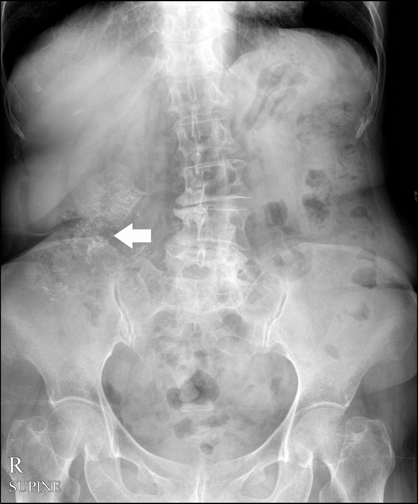
Fig. 1. Simple radiograph shows linear calcifications in the right abdominal area (arrow).
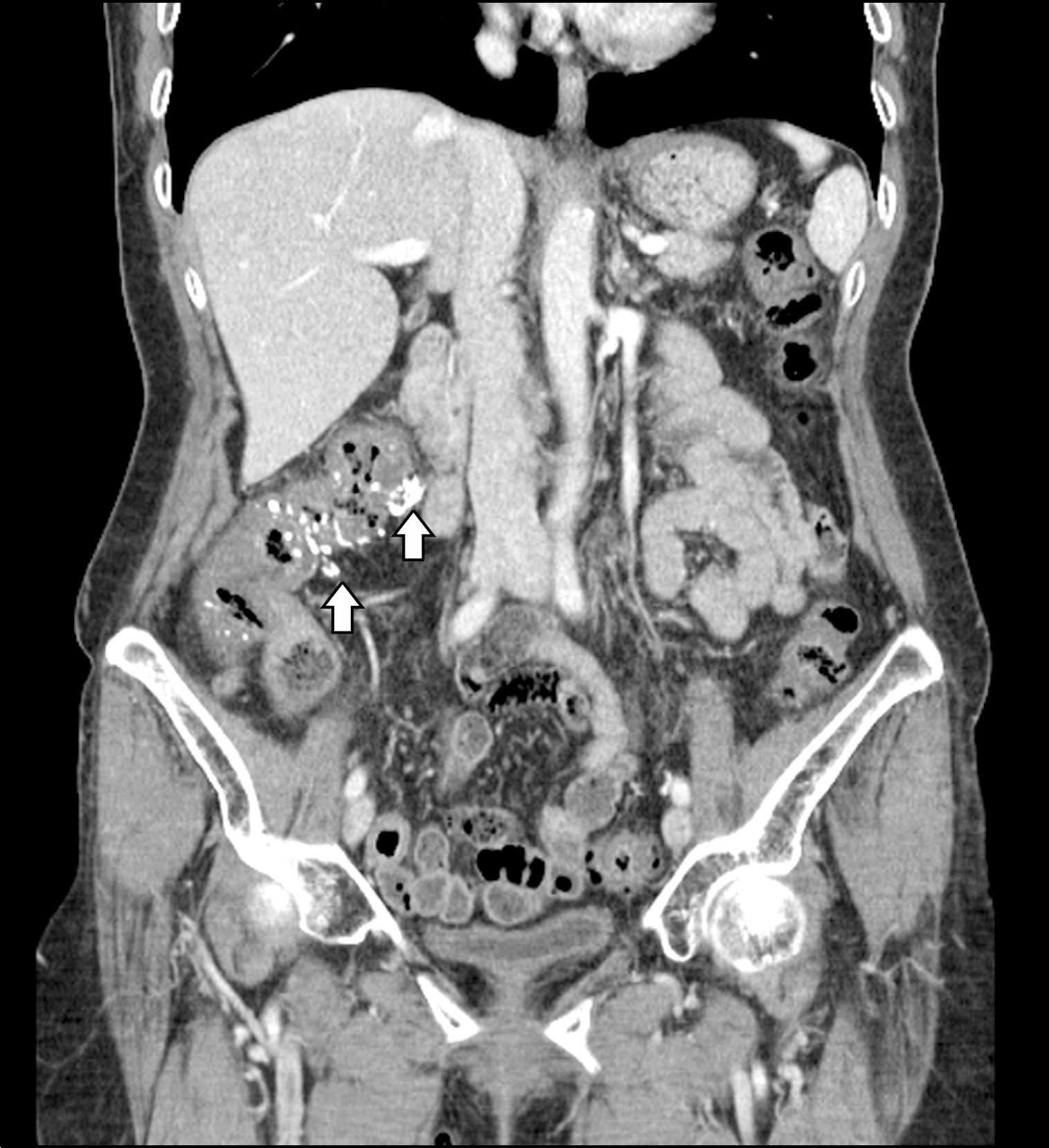
Fig. 2. Abdominal CT shows colonic wall thickenings accompanied by mural and mesenteric venous calcifications in the ascending colon (arrows).
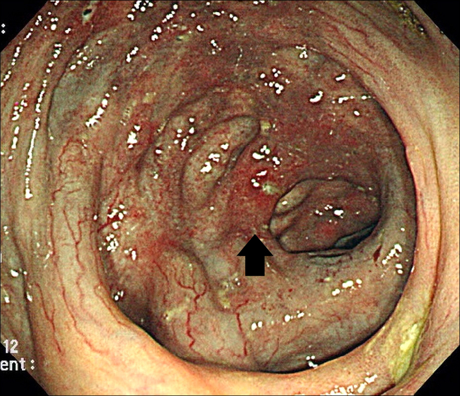
Fig. 3. Colonoscopy shows dark purple edematous mucosa with areas of luminal narrowing in the ascending colon. Multiple erosions or ulcerations with surrounding erythema are also noted (arrow).
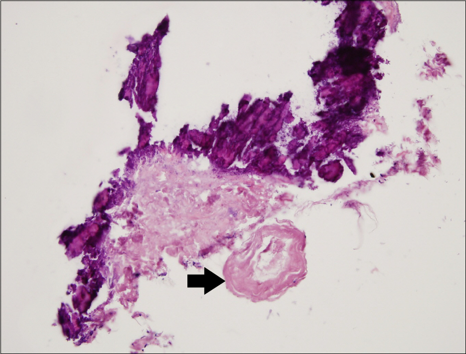
Fig. 4. Microscopic examinations. Marked thickening and calcifications of the colonic mucosal and submucosal layers are noted. Mesenteric veins show an abnormally thickened wall with hyalinization (arrow) (H&E, ×200).
Cited by 3 articles
-
Phlebosclerotic Colitis
Hosim Soh, Jaeyoung Chun
Korean J Gastroenterol. 2019;73(2):114-117. doi: 10.4166/kjg.2019.73.2.114.Phlebosclerotic Colitis in a Healthy Young Woman
Jung Kyu Park, Young Ho Sung, Sun Young Cho, Chang Yul Oh, So Hyun An
Clin Endosc. 2015;48(5):447-451. doi: 10.5946/ce.2015.48.5.447.Obstructive ileus caused by phlebosclerotic colitis
Seung Hyun Lee, Jong Wook Kim, Se Jin Park, Ju Yeol Heo, Woo Hyun Paik, Won Ki Bae, Nam-Hoon Kim, Kyung-Ah Kim, June Sung Lee
Intest Res. 2016;14(4):369-374. doi: 10.5217/ir.2016.14.4.369.
Reference
-
References
1. Macari M, Balthazar EJ. CT of bowel wall thickening: significance and pitfalls of interpretation. AJR Am J Roentgenol. 2001; 176:1105–1116.2. Iwashita A, Yao T, Schlemper RJ, et al. Mesenteric phlebo-sclerosis: a new disease entity causing ischemic colitis. Dis Colon Rectum. 2003; 46:209–220.3. Lee SH, Ha HK, Byun JY, et al. Radiological features of leiomyomatous tumors of the colon and rectum. J Comput Assist Tomogr. 2000; 24:407–412.
Article4. Lee RC, Chiang JH, Chou YH, et al. Intestinal schistosomiasis ja-ponica: CT-pathologic correlation. Radiology. 1994; 193:539–542.
Article5. Hiramatsu K, Sakata H, Horita Y, et al. Mesenteric phlebo-sclerosis associated with long-term oral intake of geniposide, an ingredient of herbal medicine. Aliment Pharmacol Ther. 2012; 36:575–586.
Article6. Chen MT, Yu SL, Yang TH. A case of phlebosclerotic colitis with involvement of the entire colon. Chang Gung Med J. 2010; 33:581–585.7. Chang KM. New histologic findings in idiopathic mesenteric phlebosclerosis: clues to its pathogenesis and etiology–prob-ably ingested toxic agent-related. J Chin Med Assoc. 2007; 70:227–235.
Article8. Kang HY, Noh R, Kim SM, Shin HD, Yun SY, Song IH. Phlebosclerotic colitis in a cirrhotic patient with portal hypertension: the first case in Korea. J Korean Med Sci. 2009; 24:1195–1199.
Article9. Song JH, Kim JI, Jung JH, et al. A case of phlebosclerotic colitis in a hemodialysis patient. Korean J Gastroenterol. 2012; 59:40–43.
Article10. Kato T, Miyazaki K, Nakamura T, Tan KY, Chiba T, Konishi F. Perforated phlebosclerotic colitis–description of a case and review of this condition. Colorectal Dis. 2010; 12:149–151.
- Full Text Links
-
- Actions
-
Cited
- CITED
-
- Close
- Share
-
- Similar articles
-
- A Case of Phlebosclerotic Colitis in a Patient of Chronic Renal Failure
- Phlebosclerotic Colitis in a Cirrhotic Patient with Portal Hypertension: The First Case in Korea
- A Case of Phlebosclerotic Colitis in a Hemodialysis Patient
- Phlebosclerotic Colitis in a Healthy Young Woman
- Phlebosclerotic Colitis in a Healthy Young Female with Long-term Herbal Medicine Use
