The Relative Contributions of the Medial Sural and Peroneal Communicating Nerves to the Sural Nerve
- Affiliations
-
- 1Department of Physical Medicine and Rehabilitation, Inha University, Inchon, Korea. jacob.kim@inha.ac.kr
- 2Department of Plastic and Reconstructive Surgery, Inha University, Inchon, Korea.
- KMID: 1715862
- DOI: http://doi.org/10.3349/ymj.2006.47.3.415
Abstract
- The medial sural cutaneous nerve (MSCN) and peroneal communicating nerve (PCN) conjoin in the calf area to form the sural nerve (SN). In previous anatomic studies, there was unresolved debate as to the main contributor to the sural nerve, and the relative contributions of MSCN and PCN had not been studied. The purpose of this study is to determine their relative neurophysiologic contributions to the SN by nerve conduction study (NCS). A total of 47 healthy subjects (25 males and 22 females, mean age 29.6+/-10.4 yrs, range 20-59 yrs) participated in the study. This study employed the orthodromic nerve conduction technique: stimulation at the ankle and recording at the mid calf (SN); specifically, we preformed stimulation at the mid calf (MSCN, PCN) and recording at 14cm proximal to the middle of the popliteal fossa (MSCN) and fibular head (PCN). The onset and peak latencies (ms) were SN 2.3+/-0.2 and 3.0+/-0.2; MSCN 2.1+/-0.2 and 2.8+/-0.2; and PCN 2.1+/-0.2 and 2.8+/-0.2. The peak-to-peak amplitudes (micro) and areas (nVsec) of the SN, MSCN, and PCN were 9.7+/-3.9, 7.0+/-4.7, and 5.0+/-3.2; and 7.2+/-2.9, 5.7+/-3.4, and 4.0+/-2.4, respectively. The side-to-side difference was not statistically significant. The main contributor to the SN was found to be the MSCN. The relative contribution ratio of the MSCN to the PCN was 1.37:1 by amplitude and 1.42:1 by area. However, in 32.9% of the subjects, the contribution of the PCN was greater than that of the MSCN.
Keyword
MeSH Terms
Figure
-

Fig. 1 Wave form markers: 1, onset latency; 2, peak latency; time from marker 1-3, duration; vertical distance from 1 (or 2)-3, peak to peak amplitude; area under the curve, from marker 1-3.

Fig. 2 Waveforms from the SN (upper trace), MSCN (middle trace), and PCN (lower trace). The values are DIST (distance), LAT (peak latency), CV (conduction velocity), and AMP (amplitude).

Fig. 3 The onset and peak latencies of the SN, MSCN and PCN. 1; the onset latency of the SN, 2; the onset latency of the MSCN, 3; the onset latency of the PCN, 4; the peak latency of the SN, 5; the peak latency of the MSCN, 6; the peak latency of the PCN.

Fig. 4 The amplitudes and areas of the SN, MSCN and PCN. 1; the amplitude of the SN, 2; the amplitude of the MSCN, 3; the amplitude of the PCN, 4; the area of the SN, 5; the area of the MSCN, 6; the area of the PCN.
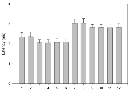
Fig. 5 The side-to-side difference for onset and peak latencies. 1; the onset latency of the SN, Rt. 2; the onset latency of the SN, Lt. 3; the onset latency of the MSCN, Rt. 4; the onset latency of the MSCN, Lt. 5; the onset latency of the PCN, Rt. 6; the onset latency of the PCN, Lt. 7 the peak latency of the SN, Rt. 8; the peak latency of the SN, Lt. 9; the peak latency of the MSCN, Rt. 10; the peak latency of the MSCN, Lt. 11; the peak latency of the PCN, Rt. 12; the peak latency of the PCN, Lt.
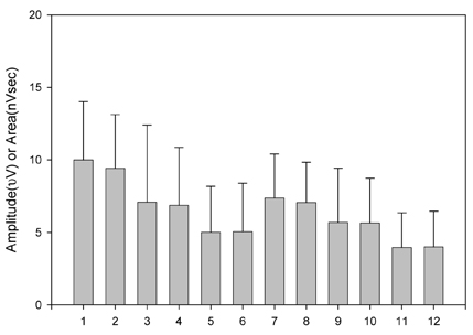
Fig. 6 The side-to-side difference for amplitudes and areas. 1; the amplitude of the SN, Rt. 2; the amplitude of the SN, Lt. 3; the amplitude of the MSCN, Rt. 4; the amplitude of the MSCN, Lt. 5; the amplitude of the PCN, Rt. 6; the amplitude of the PCN, Lt. 7; the area of the SN, Rt. 8; the area of the SN, Lt. 9; the area of the MSCN, Rt. 10; the area of the MSCN, Lt. 11; the area of the PCN, Rt. 12; the area of the PCN, Lt.

Fig. 7 The correlation of the sum amplitude with that of the SN. (r = 0.692, p < 0.01). Sum amplitude = sum amplitude of the MSCN and the PCN.

Fig. 8 The correlation of the sum area with the area of the SN. (r = 0.658, p < 0.01). Sum area = sum area of the MSCN and the PCN.
Cited by 1 articles
-
Compound Nerve Action Potential of Common Peroneal Nerve and Sural Nerve Action Potential in Common Peroneal Neuropathy
Hee Kyu Kwon, Lina Kim, Yoon Keun Park
J Korean Med Sci. 2008;23(1):117-121. doi: 10.3346/jkms.2008.23.1.117.
Reference
-
1. Aktan Ikiz ZA, Ucerler H, Bilge O. The anatomic features of the sural nerve with an emphasis on its clinical importance. Foot Ankle Int. 2005. 26:560–567.2. Berry M, Bannister LH , Standring SM. Williams PL, Bannister LH, Berry MM, Collins P, Dyson M, Dussek JE, editors. Nervous system. Gray's anatomy. 1995. 38th ed. New York: Churchill Livingstone;1286–1288.3. Coert JH, Dellon AL. Clinical implications of the surgical anatomy of the sural nerve. Plast Reconstr Surg. 1994. 94:850–855.4. De Moura W, Gilbert A. Surgical anatomy of the sural nerve. J Reconstr Microsurg. 1984. 1:31–39.5. Huelke DF. The origin of the peroneal communicating nerve in adult man. Anat Rec. 1958. 132:81–92.6. Mestdagh H, Drizenko A, Maynou C, Demondion X, Monire R. Origin and make up of the human sural nerve. Surg Radiol Anat. 2001. 23:307–312.7. Strauch B, Goldberg N, Herman CK. Sural nerve harvest: anatomy and technique. J Reconstr Microsurg. 2005. 21:133–136.8. Williams DD. A study of the human fibular communicating nerve. Anat Rec. 1954. 120:533–543.9. Liveson JA, Ma DM. Laboratory reference for clinical neurophysiology. 1992. Philadelphia: F A Davis;219–226.10. Oh SJ. Clinical electromyography: Nerve conduction studies. 1993. 2nd ed. Baltimore: Williams Wilkins;68250–251.11. Dyck PJ, Dyck PJB , Engelstad J. Dyck PJ, Thomas PK, editors. Pathologic alteration of nerves. Peripheral neuropathy. 2005. vol. 1:4th ed. Philadelphia: Elsevier Saunders;733–740.12. Oh SJ. Diagnostic usefulness and limitations of the sural nerve biopsy. Yonsei Med J. 1990. 31:1–31.13. Jobe MT, Martinez SF. Canale ST, editor. Peripheral nerve injuries. Campbell's operative orthopedics. 2003. 10th ed. Philadelphia: Mosby Co;(CD Version).14. Hill HL, Vasconez LO, Jurikiewicz MJ. Method for obtaining a sural nerve graft. Plast Resconstr Surg. 1978. 61:177–179.15. Ortiguela ME, Wood MB, Cahill DR. Anatomy of the sural nerve complex. J Hand Surg. 1987. 12A:1119–1123.16. Mahakkanukrauh P, Chomsung R. Anatomical variations of the sural nerve. Clin Anat. 2002. 15:263–266.17. Amoiridis G, Schols L, Ameridis N, Przuntek H. Motor fibers in the sural nerve of humans. Neurology. 1997. 49:1725–1728.18. Behse F, Buchthal F. Sensory action potentials and biopsy of the sural nerve in neuropathy. Brain. 1978. 101:473–493.19. Dumitru D, Amato AA, Zwarts M. Dumitru D, Amato AA, Zwarts M, editors. Nerve conduction studies. Electrodiagnostic medicine. 2002. 2nd ed. Philadelphia: Henley & Belfus;169–182.20. Buchthal F, Rosenfalk A. Evoked action potentials and conduction velocity in human sensory nerves. Brain Res. 1966. 3:1–122.21. Meythaler JM, Tuel SM, Cross LL, Reichart RT, Wertsch JJ. Electrophysiologic analysis of snap amplitude in orthodromic and antidromic studies. Electromyogr Clin Neurophysiol. 1994. 34:323–329.22. Bolton CF, Carter KM. Temperature effects on the size of human sensory compound action potentials. J Neurol Neurosurg Psychiatry. 1981. 44:407–413.23. Tilki HE, Stalberg E, Coskun M, Gungor L. Effect of heating on nerve conduction in carpal tunnel syndrome. J Clin Neurophysiol. 2004. 21:451–456.24. Cape CA. Sensory nerve action potentials of the peroneal, sural and tibial nerves. Am J Phys Med. 1971. 50:220–229.25. Buschbacher RM. Sural and saphenous 14-cm antidromic sensory nerve conduction studies. Am J Phys Med Rehabil. 2003. 82:421–426.26. Krarup C, Trojaborg W. Compound sensory action potentials evoked by tactile and by electrical stimulation in normal median and sural nerves. Muscle Nerve. 1994. 17:733–740.27. Trojaborg WT, Moon A, Andersen BB, Trojaborg NS. Sural nerve conduction parameters in normal subjects related to age, gender, temperature, and height: a reappraisal. Muscle Nerve. 1992. 15:666–671.28. Ruth A, Schulmeyer FJ, Roesch M, Woertgen C, Brawanski A. Diagnostic and therapeutic value due to suspected diagnosis, long-term complications, and indication for sural nerve biopsy. Clin Neurol Neurosurg. 2005. 107:214–217.
- Full Text Links
-
- Actions
-
Cited
- CITED
-
- Close
- Share
-
- Similar articles
-
- Sural Nerve Entrapment and Tenosynovitis of Peroneus Longus by Hypertrophied Peroneal Tubercle: A Case Report
- Anatomical Investigation of Sural Nerve and Its Contributing Nerves
- Sensitivity of Electrodiagnostic Parameters in Patients with Asymptomatic Diabetic Neuropathy
- Fascial entrapment of the sural nerve and its clinical relevance
- No Response Rates of Sensory Nerve Conduction Studies and Late Responses in Lower Limbs of Heathy Adults
