Fascial entrapment of the sural nerve and its clinical relevance
- Affiliations
-
- 1Department of Anatomy, Medical School, Aristotle University of Thessaloniki, Thessaloniki, Greece. g_paraskevas@yahoo.gr
- KMID: 1882588
- DOI: http://doi.org/10.5115/acb.2014.47.2.144
Abstract
- Sural nerve presents great topographic variability and it is responsible for sensory innervation of the posterolateral side of the distal third of the leg and lateral aspect of the foot. Entrapment of the nerve could be caused by compression due to fascial thickening, while the symptomatology includes sensory alterations and deficits at the nerve distribution area. We report a cadaveric case of a variant sural nerve that presented a distinct entrapment site. A supernumerary sensory branch was encountered originating from the common peroneal nerve, while the peroneal component of the sural nerve was observed to take a course within a fibrous fascial tunnel 3.1 cm in length that caused nerve fixation and flattening. The tension applied to the aforementioned branch was shown to worsen during passive forcible foot plantaflexion and inversion. The etiology, diagnosis and the treatment options are discussed comprehensively.
Keyword
Figure
-

Fig. 1 Entrapment of the anastomotic peroneal branch of the sural nerve. The anastomotic peroneal branch (PB) of the sural nerve was found entrapped in a fibrous fascial tunnel (FT). D, distal; L, lateral; M, medial; P, proximal; SSV, small saphenous vein.
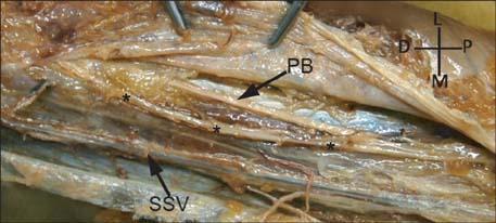
Fig. 2 Supernumerary sensory nerve originating from the common peroneal nerve. A second sensory nerve branch (*) was observed originating from the common peroneal as a separate trunk lying underneath the crural fascia and medially to the anastomotic peroneal branch (PB) of the sural nerve. D, distal; L, lateral; M, medial; P, proximal; SSV, small saphenous vein.
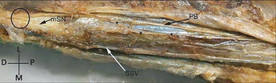
Fig. 3 Formation of the sural nerve. The sural nerve is formed by the anastomotic peroneal branch (PB) and the medial sural nerve (mSN) at the distal 1/3 of the leg. D, distal; L, lateral; M, medial; P, proximal; SSV, small saphenous vein. Circle, formation point; *, supernumerary sensory nerve.
Cited by 2 articles
-
Defining the popliteal fossa by bony landmarks and mapping of the courses of the neurovascular structures for application in popliteal fossa surgery
Kelsi Greenwood, Reinette van Zyl, Natalie Keough, Erik Hohmann
Anat Cell Biol. 2021;54(1):10-17. doi: 10.5115/acb.20.179.Extra-spinal sciatica and sciatica mimics: a scoping review
Md Abu Bakar Siddiq, Danny Clegg, Suzon Al Hasan, Johannes J Rasker
Korean J Pain. 2020;33(4):305-317. doi: 10.3344/kjp.2020.33.4.305.
Reference
-
1. Pringle RM, Protheroe K, Mukherjee SK. Entrapment neuropathy of the sural nerve. J Bone Joint Surg Br. 1974; 56B:465–468.2. Fabre T, Montero C, Gaujard E, Gervais-Dellion F, Durandeau A. Chronic calf pain in athletes due to sural nerve entrapment. A report of 18 cases. Am J Sports Med. 2000; 28:679–682.3. Pecina M, Kimpotic-Nemanic J, Markiewitz A. Tunnel syndromes. Boca Raton: CRC Press;1991. p. 105–111.4. Kosinski C. The course, mutual relations and distribution of the cutaneous nerves of the metazonal region of leg and foot. J Anat. 1926; 60(Pt 3):274–297.5. Williams DD. A study of the human fibular communicating nerve. Anat Rec. 1954; 120:533–543.6. Bruyn RP. Occupational neuropathy of the sural nerve. Ital J Neurol Sci. 1994; 15:119–120.7. Gross JA, Hamilton WJ, Swift TR. Isolated mechanical lesions of the sural nerve. Muscle Nerve. 1980; 3:248–249.8. Perlman MD. Os peroneum fracture with sural nerve entrapment neuritis. J Foot Surg. 1990; 29:119–121.9. Solomon LB, Ferris L, Tedman R, Henneberg M. Surgical anatomy of the sural and superficial fibular nerves with an emphasis on the approach to the lateral malleolus. J Anat. 2001; 199(Pt 6):717–723.10. Williams TH, Robinson AH. (iii) Entrapment neuropathies of the foot and ankle. Orthop Trauma. 2009; 23:404–411.
- Full Text Links
-
- Actions
-
Cited
- CITED
-
- Close
- Share
-
- Similar articles
-
- Entrapment of Sural Nerve in Essex-Lopresti Axial Fixation for Calcaneal Fracture: A Case Report
- Superficial Peroneal Nerve Entrapment Syndrome (A Case Report)
- Sural Nerve Entrapment by Fragments of Calcaneal Fracture (A Case Report)
- Sural Nerve Entrapment and Tenosynovitis of Peroneus Longus by Hypertrophied Peroneal Tubercle: A Case Report
- Rare case of median nerve and brachial artery entrapment by an abnormal musculo-fascial tunnel in the arm: possible cause of neurovascular compression syndrome
