Time-of-Flight Magnetic Resonance Angiography for Follow-Up of Coil Embolization with Enterprise Stent for Intracranial Aneurysm: Usefulness of Source Images
- Affiliations
-
- 1Department of Radiology, Seoul National University College of Medicine, Seoul 110-744, Korea.
- 2Department of Neurosurgery, Seoul National University College of Medicine, Seoul 110-744, Korea. hsk4428@yahoo.com
- KMID: 1711493
- DOI: http://doi.org/10.3348/kjr.2014.15.1.161
Abstract
OBJECTIVE
The aim of this study was to determine the interobserver and intermodality agreement in the interpretation of time-of-flight (TOF) MR angiography (MRA) for the follow-up of coiled intracranial aneurysms with the Enterprise stent.
MATERIALS AND METHODS
Two experienced neurointerventionists independently reviewed the follow-up MRA studies of 40 consecutive patients with 44 coiled aneurysms. All aneurysms were treated with assistance from the Enterprise stent and the radiologic follow-up intervals were greater than 6 months after the endovascular therapy. Digital subtraction angiography (DSA) served as the reference standard. The degree of aneurysm occlusion was determined by an evaluation of the maximal intensity projection (MIP) and source images (SI) of the TOF MRA. The capability of the TOF MRA to depict the residual flow within the coiled aneurysms and the stented parent arteries was compared with that of the DSA.
RESULTS
DSA showed stable occlusions in 25 aneurysms, minor recanalization in 8, and major recanalization in 11. Comparisons between the TOF MRA and conventional angiography showed that the MIP plus SI had almost perfect agreement (kappa = 0.892, range 0.767 to 1.000) and had better agreement than with the MIP images only (kappa = 0.598, range 0.370 to 0.826). In-stent stenosis of more than 33% was observed in 5 cases. Both MIP and SI of the MRA showed poor depiction of in-stent stenosis compared with the DSA.
CONCLUSION
TOF MRA seemed to be reliable in screening for aneurysm recurrence after coil embolization with Enterprise stent assistance, especially in the evaluation of the SI, in addition to MIP images in the TOF MRA.
Keyword
MeSH Terms
-
Angiography, Digital Subtraction/methods
Cerebral Angiography/methods
Embolization, Therapeutic/instrumentation/*methods
Female
Follow-Up Studies
Humans
Intracranial Aneurysm/diagnosis/radiography/*therapy
Magnetic Resonance Angiography/*methods
Male
Middle Aged
Observer Variation
Recurrence
Reference Standards
*Stents
Figure
-

Fig. 1 Examples of angiographic interpretation.Degree of coiled aneurysmal stability was classified as stable occlusion (A), minor recanalization (B), and major recanalization (C).
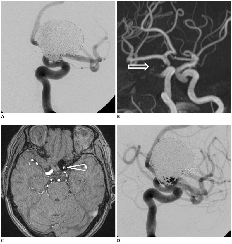
Fig. 2 Exemplary case of major recanalization.A. Cerebral angiography after coil embolization using Enterprise stent for paraclinoid internal carotid artery aneurysms showed neck remnant with small size. B. In TOF MRA performed at 6 month follow-up examination, MIP image did not show any flow within coiled aneurysm (arrow indicates stented parent artery). C. However, some flow within aneurysmal sac was suspected in source image (arrowhead indicates recanalized portion). D. Conventional angiography showed aneurysm to be visibly recanalized. TOF = time-of-flight, MRA = MR angiography
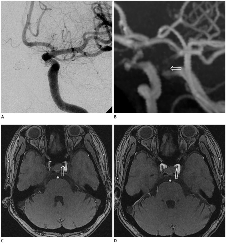
Fig. 3 Exemplary case of mismatched interpretation.One-year follow-up angiography showed major recanalization of coiled aneurysm in distal internal carotid artery (A). However, MIP (B) and source image (C) of TOF MRA failed to show any flow within aneurysm (arrows indicate coiled aneurysm). This was in contrast to source image of pre-embolization MRA (D) (arrowhead indicates untreated aneurysm). TOF = time-of-flight, MRA = MR angiography, MIP = maximal intensity projection
Cited by 4 articles
-
De Novo Intracranial Aneurysms Detected on Imaging Follow-Up of Coiled Aneurysms in a Korean Population
Eung Koo Yeon, Young Dae Cho, Dong Hyun Yoo, Su Hwan Lee, Hyun-Seung Kang, Won Sang Cho, Jeong Eun Kim, Moon Hee Han
Korean J Radiol. 2019;20(9):1390-1398. doi: 10.3348/kjr.2018.0914.Accelerated Time-of-Flight Magnetic Resonance Angiography with Sparse Undersampling and Iterative Reconstruction for the Evaluation of Intracranial Arteries
Hehan Tang, Na Hu, Yuan Yuan, Chunchao Xia, Xiumin Liu, Panli Zuo, Aurelien F. Stalder, Michaela Schmidt, Xiaoyue Zhou, Bin Song, Jiayu Sun
Korean J Radiol. 2019;20(2):265-274. doi: 10.3348/kjr.2017.0634.Microcatheter Stabilization Technique Using Partially Inflated Balloon for Coil Embolization of Paraclinoid Aneurysms
Yunsun Song, Boseong Kwon, Abdulrahman Hamad Al-abdulwahhab, Ricky Gusanto Kurniawan, Dae Chul Suh
Neurointervention. 2021;16(2):132-140. doi: 10.5469/neuroint.2021.00185.Does a Low-wall Coverage Stent Have a Flow Diverting Effect in Small Aneurysms?
Hairi Liu, Jooae Choe, Seung Chul Jung, Yunsun Song, Ku Hyun Yang, Kye Jin Park, Hae Won Goo, Won Hyong Park, Dae Chul Suh
Neurointervention. 2015;10(2):89-93. doi: 10.5469/neuroint.2015.10.2.89.
Reference
-
1. Molyneux A, Kerr R, Stratton I, Sandercock P, Clarke M, Shrimpton J, et al. International Subarachnoid Aneurysm Trial (ISAT) of neurosurgical clipping versus endovascular coiling in 2143 patients with ruptured intracranial aneurysms: a randomised trial. Lancet. 2002; 360:1267–1274. PMID: 12414200.
Article2. Lavoie P, Gariépy JL, Milot G, Jodoin S, Bédard F, Trottier F, et al. Residual flow after cerebral aneurysm coil occlusion: diagnostic accuracy of MR angiography. Stroke. 2012; 43:740–746. PMID: 22267824.3. Piotin M, Blanc R, Spelle L, Mounayer C, Piantino R, Schmidt PJ, et al. Stent-assisted coiling of intracranial aneurysms: clinical and angiographic results in 216 consecutive aneurysms. Stroke. 2010; 41:110–115. PMID: 19959540.4. Choi JW, Roh HG, Moon WJ, Kim NR, Moon SG, Kang CH, et al. Time-resolved 3D contrast-enhanced MRA on 3.0T: a non-invasive follow-up technique after stent-assisted coil embolization of the intracranial aneurysm. Korean J Radiol. 2011; 12:662–670. PMID: 22043147.
Article5. Takayama K, Taoka T, Nakagawa H, Myouchin K, Wada T, Sakamoto M, et al. Usefulness of contrast-enhanced magnetic resonance angiography for follow-up of coil embolization with the enterprise stent for cerebral aneurysms. J Comput Assist Tomogr. 2011; 35:568–572. PMID: 21926851.
Article6. Choi JW, Roh HG, Moon WJ, Chun YI, Kang CH. Optimization of MR Parameters of 3D TOF-MRA for Various Intracranial Stents at 3.0T MRI. Neurointervention. 2011; 6:71–77. PMID: 22125752.
Article7. Raymond J, Guilbert F, Weill A, Georganos SA, Juravsky L, Lambert A, et al. Long-term angiographic recurrences after selective endovascular treatment of aneurysms with detachable coils. Stroke. 2003; 34:1398–1403. PMID: 12775880.
Article8. Kang HS, Han MH, Kwon BJ, Kwon OK, Kim SH, Choi SH, et al. Short-term outcome of intracranial aneurysms treated with polyglycolic acid/lactide copolymer-coated coils compared to historical controls treated with bare platinum coils: a single-center experience. AJNR Am J Neuroradiol. 2005; 26:1921–1928. PMID: 16155135.9. Lee SJ, Cho YD, Kang HS, Kim JE, Han MH. Coil embolization using the self-expandable closed-cell stent for intracranial saccular aneurysm: a single-center experience of 289 consecutive aneurysms. Clin Radiol. 2013; 68:256–263. PMID: 23017739.
Article10. Kovács A, Möhlenbruch M, Hadizadeh DR, Seifert M, Greschus S, Clusmann H, et al. Noninvasive imaging after stent-assisted coiling of intracranial aneurysms: comparison of 3-T magnetic resonance imaging and 64-row multidetector computed tomography--a pilot study. J Comput Assist Tomogr. 2011; 35:573–582. PMID: 21926852.11. Pierot L, Portefaix C, Boulin A, Gauvrit JY. Follow-up of coiled intracranial aneurysms: comparison of 3D time-of-flight and contrast-enhanced magnetic resonance angiography at 3T in a large, prospective series. Eur Radiol. 2012; 22:2255–2263. PMID: 22569997.
Article12. Schaafsma JD, Velthuis BK, Majoie CB, van den Berg R, Brouwer PA, Barkhof F, et al. Intracranial aneurysms treated with coil placement: test characteristics of follow-up MR angiography--multicenter study. Radiology. 2010; 256:209–218. PMID: 20505063.
Article13. Plank CM, Wolf F, Langenberger H, Weber M, Beitzke D, Stadler A, et al. Improved detection of in-stent restenosis by blood pool agent-enhanced, high-resolution, steady-state magnetic resonance angiography. Eur Radiol. 2011; 21:2158–2165. PMID: 21556908.
Article14. Agid R, Willinsky RA, Lee SK, Terbrugge KG, Farb RI. Characterization of aneurysm remnants after endovascular treatment: contrast-enhanced MR angiography versus catheter digital subtraction angiography. AJNR Am J Neuroradiol. 2008; 29:1570–1574. PMID: 18499789.
Article15. Higashida RT, Halbach VV, Dowd CF, Juravsky L, Meagher S. Initial clinical experience with a new self-expanding nitinol stent for the treatment of intracranial cerebral aneurysms: the Cordis Enterprise stent. AJNR Am J Neuroradiol. 2005; 26:1751–1756. PMID: 16091525.16. Weber W, Bendszus M, Kis B, Boulanger T, Solymosi L, Kühne D. A new self-expanding nitinol stent (Enterprise) for the treatment of wide-necked intracranial aneurysms: initial clinical and angiographic results in 31 aneurysms. Neuroradiology. 2007; 49:555–561. PMID: 17476494.
Article17. Mocco J, Fargen KM, Albuquerque FC, Bendok BR, Boulos AS, Carpenter JS, et al. Delayed thrombosis or stenosis following enterprise-assisted stent-coiling: is it safe? Midterm results of the interstate collaboration of enterprise stent coiling. Neurosurgery. 2011; 69:908–913. discussion 913-914. PMID: 21670718.
Article18. Gauvrit JY, Leclerc X, Caron S, Taschner CA, Lejeune JP, Pruvo JP. Intracranial aneurysms treated with Guglielmi detachable coils: imaging follow-up with contrast-enhanced MR angiography. Stroke. 2006; 37:1033–1037. PMID: 16528000.19. Seok JH, Choi HS, Jung SL, Ahn KJ, Kim MJ, Shin YS, et al. Artificial luminal narrowing on contrast-enhanced magnetic resonance angiograms on an occasion of stent-assisted coiling of intracranial aneurysm: in vitro comparison using two different stents with variable imaging parameters. Korean J Radiol. 2012; 13:550–556. PMID: 22977321.
- Full Text Links
-
- Actions
-
Cited
- CITED
-
- Close
- Share
-
- Similar articles
-
- Time-Resolved 3D Contrast-Enhanced MRA on 3.0T: a Non-Invasive Follow-Up Technique after Stent-Assisted Coil Embolization of the Intracranial Aneurysm
- Stent-Assisted Coil Embolization for the Proximal Middle Cerebral Artery Fusiform Aneurysm
- Usefulness of Silent MRA for Evaluation of Aneurysm after Stent-Assisted Coil Embolization
- Blood Blister-like Aneurysm Treated with Coil Embolization without Stent Assistance
- In-Stent Stenosis of Stent Assisted Endovascular Treatment on Intracranial Complex Aneurysms
