J Korean Soc Magn Reson Med.
2014 Mar;18(1):1-6. 10.13104/jksmrm.2014.18.1.1.
Estimation of T2* Relaxation Times for the Glandular Tissue and Fat of Breast at 3T MRI System
- Affiliations
-
- 1Department of Radiology, Kyung Hee University Hospital at Gangdong, College of Medicine, Kyung Hee University, Seoul, Korea. ghjahng@gmail.com
- 2Department of Biomedical Engineering, College of Electronics and Information Kyung Hee University, Yongin, Korea.
- 3Graduate College of Medicine, Kyung Hee University, Seoul, Korea.
- KMID: 1683774
- DOI: http://doi.org/10.13104/jksmrm.2014.18.1.1
Abstract
- PURPOSE
T2* relaxation time which includes susceptibility information represents unique feature of tissue. The objective of this study was to investigate T2* relaxation times of the normal glandular tissue and fat of breast using a 3T MRI system.
MATERIALS AND METHODS
Seven-echo MR Images were acquired from 52 female subjects (age 49 +/- 12 years; range, 25 to 75) using a three-dimensional (3D) gradient-echo sequence. Echo times were between 2.28 ms to 25.72 ms in 3.91 ms steps. Voxel-based T2* relaxation times and R2* relaxation rate maps were calculated by using the linear curve fitting for each subject. The 3D regions-of-interest (ROI) of the normal glandular tissue and fat were drawn on the longest echo-time image to obtain T2* and R2* values. Mean values of those parameters were calculated over all subjects.
RESULTS
The 3D ROI sizes were 4818 +/- 4679 voxels and 1455 +/- 785 voxels for the normal glandular tissue and fat, respectively. The mean T2* values were 22.40 +/- 5.61 ms and 36.36 +/- 8.77 ms for normal glandular tissue and fat, respectively. The mean R2* values were 0.0524 +/- 0.0134/ms and 0.0297 +/- 0.0069/ms for the normal glandular tissue and fat, respectively.
CONCLUSION
T2* and R2* values were measured from human breast tissues. T2* of the normal glandular tissue was shorter than that of fat. Measurement of T2* relaxation time could be important to understand susceptibility effects in the breast cancer and the normal tissue.
Keyword
Figure
-
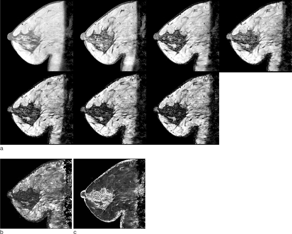
Fig. 1 Representative images obtained and calculated from a subject (71 year-old woman). a. Sagittal slices obtained using the three-dimensional multi-echo gradient-echo sequence. Seven echo times (TE) were from 2.28 ms to 25.72 ms in 3.91 ms steps. b. T2* relaxometry maps c. R2* relaxometry maps
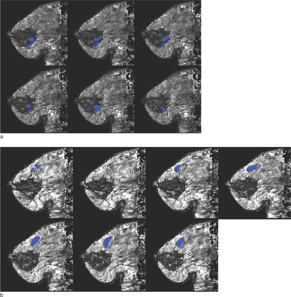
Fig. 2 Representative regions-of-interest overlayed in T2* map in the same subject of Fig. 1. a. Fibroglandular tissue b. Fat
Reference
-
1. Cheng HL, Stikov N, Ghugre NR, Wright GA. Practical medical applications of quantitative MR relaxometry. J Magn Reson Imaging. 2012; 36:805–824.2. Carneiro AAO, Vilela GR, de Araujo DB, Baffa O. MRI relaxometry: methods and applications. Braz J Phys. 2006; 36:9–15.3. Chavhan GB, Babyn PS, Thomas B, Shroff MM, Haacke EM. Principles, techniques, and applications of T2*-based MR imaging and its special applications. Radiographics. 2009; 29:1433–1449.4. Rakow-Penner R, Daniel B, Yu H, Sawyer-Glover A, Glover GH. Relaxation times of breast tissue at 1.5T and 3T measured using IDEAL. J Magn Reson Imaging. 2006; 23:87–91.5. Edden RA, Smith SA, Barker PB. Longitudinal and multi-echo transverse relaxation times of normal breast tissue at 3 Tesla. J Magn Reson Imaging. 2010; 32:982–987.6. Christen T, Bolar DS, Zaharchuk G. Imaging brain oxygenation with MRI using blood oxygenation approaches: methods, validation, and clinical applications. AJNR Am J Neuroradiol. 2013; 34:1113–1123.7. Tsushima Y, Endo K. Hypointensities in the brain on T2*-weighted gradient-echo magnetic resonance imaging. Curr Probl Diagn Radiol. 2006; 35:140–150.8. Uludag K, Dubowitz DJ, Buxton RB. Basic principles of functional MRI. In : Edelman RR, Hesselink JR, Zlatkin MB, Crues JV, editors. Clinical magnetic resonance imaging. 3rd ed. Philadelphia, Pa: Saunders Elsevier;2006. p. 249–287.9. Hendrick RE. Image contrast and noise. In : Stark DD, Bradley WG, editors. Magnetic resonance imaging. 3rd ed. St Louis, Mo: Mosby;1999. p. 43–68.10. Haacke EM, Tkach JA, Parrish TB. Reduction of T2* dephasing in gradient field-echo imaging. Radiology. 1989; 170:457–462.11. Frahm J, Haenicke W. Rapid scan techniques. In : Stark DD, Bradley WG, editors. Magnetic resonance imaging. 3rd ed. St Louis, Mo: Mosby;1999. p. 87–124.12. Busk M, Horsman MR. Relevance of hypoxia in radiation oncology: pathophysiology, tumor biology and implications for treatment. Q J Nucl Med Mol Imaging. 2013; 57:219–234.
- Full Text Links
-
- Actions
-
Cited
- CITED
-
- Close
- Share
-
- Similar articles
-
- Phantom-Validated Reference Values of Myocardial Mapping and Extracellular Volume at 3T in Healthy Koreans
- Estimation of T2* Relaxation Time of Breast Cancer: Correlation with Clinical, Imaging and Pathological Features
- A study on magnetic relaxation times of various organs and body fluids using superconducting magnetic resonanceimaging system
- High field strength magnetic resonance imaging in children
- Improvement of Fat Suppression and Artifact Reduction Using IDEAL Technique in Head and Neck MRI at 3T
