The Usefulness of the Ivy Sign on Fluid-Attenuated Intensity Recovery Images in Improved Brain Hemodynamic Changes after Superficial Temporal Artery-Middle Cerebral Artery Anastomosis in Adult Patients with Moyamoya Disease
- Affiliations
-
- 1Department of Neurosurgery, Eulji University Hospital, College of Medicine, Eulji University, Daejeon, Korea. neurocsy@eulji.ac.kr
- KMID: 1499335
- DOI: http://doi.org/10.3340/jkns.2013.54.4.302
Abstract
OBJECTIVE
MR perfusion and single photon emission computerized tomography (SPECT) are well known imaging studies to evaluate hemodynamic change between prior to and following superficial temporal artery (STA)-middle cerebral artery (MCA) anastomosis in moyamoya disease. But their side effects and invasiveness make discomfort to patients. We evaluated the ivy sign on MR fluid attenuated inversion recovery (FLAIR) images in adult patients with moyamoya disease and compared it with result of SPECT and MR perfusion images.
METHODS
We enrolled twelve patients (thirteen cases) who were diagnosed with moyamoya disease and underwent STA-MCA anastomosis at our medical institution during a period ranging from September of 2010 to December of 2012. The presence of the ivy sign on MR FLAIR images was classified as Negative (0), Minimal (1), and Positive (2). Regions were classified into four territories: the anterior cerebral artery (ACA), the anterior MCA, the posterior MCA and the posterior cerebral artery.
RESULTS
Ivy signs on preoperative and postoperative MR FLAIR were improved (8 and 4 in the ACA regions, 13 and 4 in the anterior MCA regions and 19 and 9 in the posterior MCA regions). Like this result, the cerebrovascular reserve (CVR) on SPECT was significantly increased in the sum of CVR in same regions after STA-MCA anastomosis.
CONCLUSION
After STA-MCA anastomosis, ivy signs were decreased in the cerebral hemisphere. As compared with conventional diagnostic modalities such as SPECT and MR perfusion images, the ivy sign on MR FLAIR is considered as a useful indicator in detecting brain hemodynamic changes between preoperatively and postoperatively in adult moyamoya patients.
Keyword
MeSH Terms
Figure
-
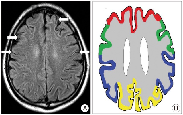
Fig. 1 The ivy sign is referred to as leptomeningeal high-signal intensity on FLAIR images along cerebral sulci. White arrows indicate ivy signs on bilateral hemispheres of moyamoya patient (A). Regions on bilateral hemispheres were classified into four territories16) (AJNR Am J Neuroradiol 20 : 336-343, 1999) : Red line - ACA, Green line - the anterior-MCA, Pupple line - the posterior MCA, Yellow line - PCA (B). ACA : Anterior cerebral artery, MCA : Middle cerebral artery, PCA : posterior cerebral artery, FLAIR : fluid-attenuated inversion recovery.
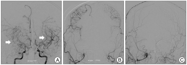
Fig. 2 A 36-year-old man with moyamoya disease who got the bilateral STA-MCA anastomosis surgery in our hospital. In the preoperative TFCA image, white arrows indicate moyamoya vessels on bilateral ICA (A). No vascular supply to intracranial region via ECA (B). STA-MCA anastomosis was done on bilateral hemisphere and well intracranial blood flow was checked from STA to MCA via anastomosis site in the postoperative TFCA image (C). TFCA : transfemoral cerebral angiography, STA : superficial temporal artery, ICA : internal carotid artery, ECA : external carotid artery, MCA : middle cerebral artery.

Fig. 3 Same patient with Fig. 2. Ivy sign was checked on bilateral hemispheres by indicating white arrows (A) and compared preoperative MR with postoperative MR FLAIR image, ivy sign was disappeared on bilateral hemispheres by indicating white arrows (B). Like MR FLAIR images, compared with preoperative SPECT and postoperative SPECT, CVR was increased on bilateral hemisphere before surgical treatment by indicating white arrowheads (C and D). SPECT : single photon emission computerized tomography, CVR : cerebrovascular reserve, FLAIR : fluid-attenuated inversion recovery.
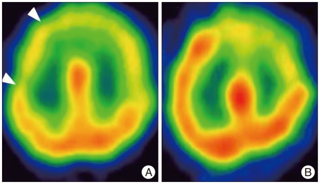
Fig. 4 A 64-year-old woman with moyamoya disease. White arrowheads indicate the decreased CVR with SPECT images (A). The patient got the surgery, and decreased CVR was normalization on SPECT images after 1 month (B). CVR : cerebrovascular reserve, SPECT : single photon emission computerized tomography.

Fig. 5 Same patient with Fig. 4. We compared ivy sign on MR FLAIR image with MTT on MR perfusion image. Preoperatively, white arrows indicate the ivy signs on MR FLAIR image (A) and those were decreased on MR FLAIR images after surgery (B). MTT prolongation was observed on MR perfusion images in the right MCA territory in the preoperative state by indicating white arrows, and after surgery, MTT prolongation was improved on MR perfusion images like ivy signs on MR FLAIR images (C and D). MTT : mean transit time, FLAIR : fluid-attenuated inversion recovery.
Reference
-
1. Baba T, Houkin K, Kuroda S. Novel epidemiological features of moyamoya disease. J Neurol Neurosurg Psychiatry. 2008; 79:900–904. PMID: 18077479.
Article2. Cosnard G, Duprez T, Grandin C, Smith AM, Munier T, Peeters A. Fast FLAIR sequence for detecting, major vascular abnormalities during the hyperacute phase of stroke : a comparison with MR angiography. Neuroradiology. 1999; 41:342–346. PMID: 10379591.
Article3. Fujimura M, Mugikura S, Shimizu H, Tominaga T. Diagnostic value of perfusion-weighted MRI for evaluating postoperative alteration of cerebral hemodynamics following STA-MCA anastomosis in patients with moyamoya disease. No Shinkei Geka. 2006; 34:801–809. PMID: 16910493.4. Fujiwara H, Momoshima S, Kuribayashi S. Leptomeningeal high signal intensity (ivy sign) on fluid-attenuated inversion-recovery (FLAIR) MR images in moyamoya disease. Eur J Radiol. 2005; 55:224–230. PMID: 16036151.
Article5. Gauvrit JY, Leclerc X, Girot M, Cordonnier C, Sotoares G, Henon H, et al. Fluid-attenuated inversion recovery (FLAIR) sequences for the assessment of acute stroke : inter observer and intertechnique reproducibility. J Neurol. 2006; 253:631–635. PMID: 16362529.
Article6. Ideguchi R, Morikawa M, Enokizono M, Ogawa Y, Nagata I, Uetani M. Ivy signs on FLAIR images before and after STA-MCA anastomosis in patients with Moyamoya disease. Acta Radiol. 2011; 52:291–296. PMID: 21498365.
Article7. Iida H, Itoh H, Nakazawa M, Hatazawa J, Nishimura H, Onishi Y, et al. Quantitative mapping of regional cerebral blood flow using iodine-123-IMP and SPECT. J Nucl Med. 1994; 35:2019–2030. PMID: 7989987.8. Kawamura Y, Ashizaki M, Saida S, Sugimoto H. Usefulness of rate of increase in SPECT counts in one-daymethod of N-isopropyl-4-iodoamphetamine [123I] SPECT studies at rest and after acetazolamide challenge using amethod for estimating time-dependent distribution at rest. Ann Nucl Med. 2008; 22:457–463. PMID: 18600426.
Article9. Kawashima M, Noguchi T, Takase Y, Nakahara Y, Matsushima T. Decrease in leptomeningeal ivy sign on fluid-attenuated inversion recovery images after cerebral revascularization in patients with moyamoya disease. AJNR Am J Neuroradiol. 2010; 31:1713–1718. PMID: 20466798.
Article10. Kawashima M, Noguchi T, Takase Y, Ootsuka T, Kido N, Matsushima T. Unilateral hemispheric proliferation of ivy sign on FLAIR image in Moyamoya disease correlates highly with ipsilateral hemispheric decrease of cerebrovascular reserve. AJNR Am J Neuroradiol. 2009; 30:1709–1716. PMID: 19713323.
Article11. Komiyama M, Nakajima H, Nishikawa M, Yasui T, Kitano S, Sakamoto H. Leptomeningeal contrast enhancement in moyamoya : its potential role in postoperative assessment of circulation through the bypass. Neuroradiology. 2001; 43:17–23. PMID: 11214642.
Article12. Lee SK, Kim DI, Jeong EK, Kim SY, Kim SH, In YK, et al. Postoperative evaluation of moyamoya disease with perfusion-weighted MR imaging : initial experience. AJNR Am J Neuroradiol. 2003; 24:741–747. PMID: 12695215.13. Maeda M, Tsuchida C. "Ivy sign" on fluid-attenuated inversion-recovery images in childhood moyamoya disease. AJNR Am J Neuroradiol. 1999; 20:1836–1838. PMID: 10588105.14. Maeda M, Yamamoto T, Daimon S, Sakuma H, Takeda K. Arterial hyperintensity on fast fluid-attenuated inversion recovery images : a subtle finding for hyperacute stroke undetected by diffusion-weighted MR imaging. AJNR Am J Neuroradiol. 2001; 22:632–636. PMID: 11290469.15. Mori N, Mugikura S, Higano S, Kaneta T, Fujimura M, Umetsu A, et al. The leptomeningeal "ivy sign" on fluid-attenuated inversion recovery MR imaging in Moyamoya disease : a sign of decreased cerebral vascular reserve? AJNR Am J Neuroradiol. 2009; 30:930–935. PMID: 19246527.
Article16. Mugikura S, Takahashi S, Higano S, Shirane R, Kurihara N, Furuta S, et al. The relationship between cerebral infarction and angiographic characteristics in childhood moyamoya disease. AJNR Am J Neuroradiol. 1999; 20:336–343. PMID: 10094366.17. Narisawa A, Fujimura M, Tominaga T. Efficacy of the revascularization surgery for adult-onset moyamoya disease with the progression of cerebrovascular lesions. Clin Neurol Neurosurg. 2009; 111:123–126. PMID: 18995956.
Article18. Noguchi K, Ogawa T, Inugami A, Fujita H, Hatazawa J, Shimosegawa E, et al. MRI of acute cerebral infarction : a comparison of FLAIR and T2-weighted fast spin-echo imaging. Neuroradiology. 1997; 39:406–410. PMID: 9225318.
Article19. Ohta T, Tanaka H, Kuroiwa T. Diffuse leptomeningeal enhancement, "ivy sign," in magnetic resonance images of moyamoya disease in childhood. Case report. Neurosurgery. 1995; 37:1009–1012. PMID: 8559324.
Article20. Ringelstein EB, Sievers C, Ecker S, Schneider PA, Otis SM. Noninvasive assessment of CO2-induced cerebral vasomotor response in normal individuals and patients with internal carotid artery occlusions. Stroke. 1988; 19:963–969. PMID: 3135641.
Article21. Ringelstein EB, Van Eyck S, Mertens I. Evaluation of cerebral vasomotor reactivity by various vasodilating stimuli : comparison of CO2 to acetazolamide. J Cereb Blood Flow Metab. 1992; 12:162–168. PMID: 1727137.
Article
- Full Text Links
-
- Actions
-
Cited
- CITED
-
- Close
- Share
-
- Similar articles
-
- A Middle Cerebral Artery AneurysmOriginating Near the Site of Anastomosis after Superficial Temporal Artery-Middle Cerebral Artery Bypass: Case Report
- Ivy Sign on Fluid-Attenuated Inversion Recovery Images in Moyamoya Disease: Correlation with Clinical Severity and Old Brain Lesions
- Clinical Analysis of Surgically Treated Moyamoya Diseases
- Development of Brain Infarction after Extracranial-Intracranial Bypass Surgery in a Patient with Moyamoya Disease: A case report
- Long-Term Follow-up Study after Superficial Temporal Artery-Middle Cerebral Artery Anastomosis plus Encephalomyosynangiosis for Moyamoya Disease
