Analysis of Measurement Accuracy for Craniovertebral Junction Pathology : Most Reliable Method for Cephalometric Analysis
- Affiliations
-
- 1Department of Neurosurgery, St. Vincent's Hospital, The Catholic University of Korea, Suwon, Korea. jatagi15@paran.com
- KMID: 1499331
- DOI: http://doi.org/10.3340/jkns.2013.54.4.275
Abstract
OBJECTIVE
This study was designed to determine the most reliable cephalometric measurement technique in the normal population and patients with basilar invagination (BI).
METHODS
Twenty-two lateral radiographs of BI patients and 25 lateral cervical radiographs of the age, sex-matched normal population were selected and measured on two separate occasions by three spine surgeons using six different measurements. Statistical analysis including intraclass correlation coefficient (ICC) was carried out using the SPSS software (V. 12.0).
RESULTS
Redlund-Johnell and Modified (M)-Ranawat had a highest ICC score in both the normal and BI groups in the inter-observer study. The M-Ranawat method (0.83) had a highest ICC score in the normal group, and the Redlund-Johenll method (0.80) had a highest ICC score in the BI group in the intra-observer test. The McGregor line had a lowest ICC score and a poor ICC grade in both groups in the intra-observer study. Generally, the measurement method using the odontoid process did not produce consistent results due to inter and intra-observer differences in determining the position of the odontoid tip. Opisthion and caudal point of the occipital midline curve are somewhat ambiguous landmarks, which induce variable ICC scores.
CONCLUSION
On the contrary to other studies, Ranawat method had a lower ICC score in the inter-observer study. C2 end-plate and C1 arch can be the most reliable anatomical landmarks.
Keyword
MeSH Terms
Figure
-
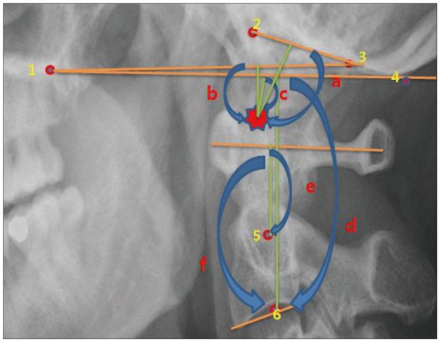
Fig. 1 Relevant landmarks and six-different measurements. 1 : Hard palate, 2 : Basion, 3 : Opisthion, 4 : The most caudal point on the midline occipital curve, 5 : Center of the second cervical pedicle, 6 : Midpoint of the caudal margin of the second cervical vertebra body, a : McRae line, b : Chamberlain line c, : McGregor line, d : Redlund-Johnell method, e : Ranawat method, f : Modified-Ranawat method, Asterion : odontoid tip.
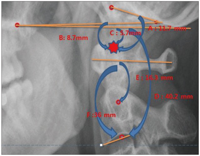
Fig. 2 Measuring the six-differents methods in normal patient. A : McRae method, B : Chamberlain method, C : McGregor method, D : Redlund-Johnell method, E : Ranawat method, F : Modified-Ranawat method, Asterion : odontoid tip.
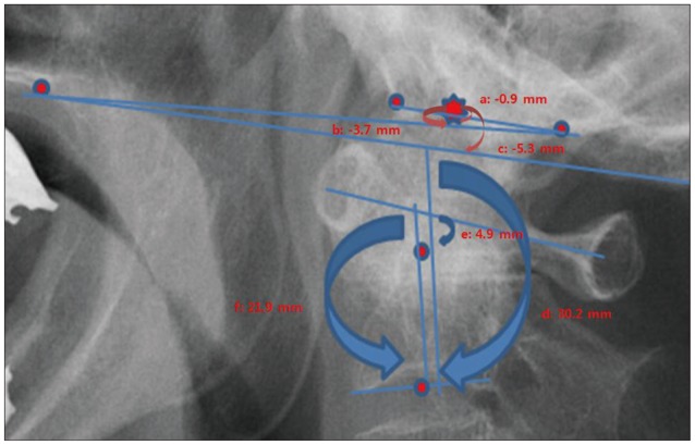
Fig. 3 Measuring the six-differents methods in basilar invagination patient. a : McRae method, b : Chamberlain method, c : McGregor method, d : Redlund-Johnell method, e : Ranawat method, f : Modified-Ranawat method, Asterion : odontoid tip.

Fig. 4 This graphs show the intraclass correlation coefficient score (original ICC score times one hundred) with inter-observer study. BI : basilar invagination.
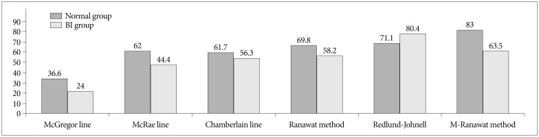
Fig. 5 This graphs shows the intraclass correlation coefficient score (original ICC score times one hundred) with intra-observer study. BI : basilar invagination.
Reference
-
1. Chamberlain WE. Basilar Impression (Platybasia) : A Bizarre Developmental Anomaly of the Occipital Bone and Upper Cervical Spine with Striking and Misleading Neurologic Manifestations. Yale J Biol Med. 1939; 11:487–496. PMID: 21433841.2. Clark CR, Goetz DD, Menezes AH. Arthrodesis of the cervical spine in rheumatoid arthritis. J Bone Joint Surg Am. 1989; 71:381–392. PMID: 2925711.
Article3. Dziurzynski K, Anderson PA, Bean DB, Choi J, Leverson GE, Marin RL, et al. A blinded assessment of radiographic criteria for atlanto-occipital dislocation. Spine (Phila Pa 1976). 2005; 30:1427–1432. PMID: 15959373.
Article4. Kauppi M, Sakaguchi M, Konttinen YT, Hamalainen M. A new method of screening for vertical atlantoaxial dislocation. J Rheumatol. 1990; 17:167–172. PMID: 2181126.5. Kawaida H, Sakou T, Morizono Y. Vertical settling in rheumatoid arthritis. Diagnostic value of the Ranawat and Redlund-Johnell methods. Clin Orthop Relat Res. 1989; 239:128–135. PMID: 2912612.6. Kwong Y, Rao N, Latief K. Craniometric measurements in the assessment of craniovertebral settling : are they still relevant in the age of cross-sectional imaging? AJR Am J Roentgenol. 2011; 196:W421–W425. PMID: 21427306.7. McGreger M. The significance of certain measurements of the skull in the diagnosis of basilar impression. Br J Radiol. 1948; 21:171–181. PMID: 18912046.
Article8. McRae DL, Barnum AS. Occipitalization of the atlas. Am J Roentgenol Radium Ther Nucl Med. 1953; 70:23–46.9. Mikulowski P, Wollheim FA, Rotmil P, Olsen I. Sudden death in rheumatoid arthritis with atlanto-axial dislocation. Acta Med Scand. 1975; 198:445–451. PMID: 1211212.
Article10. Nagayoshi R, Ijiri K, Takenouchi T, Taketomi E, Sakakima H, Komiya S. Evaluation of occipitocervical subluxation in rheumatoid arthritis patients, using coronal-view reconstructive computed tomography. Spine (Phila Pa 1976). 2009; 34:E879–E881. PMID: 19910756.
Article11. Parish DC, Clark JA, Liebowitz SM, Hicks WC. Sudden death in rheumatoid arthritis from vertical subluxation of the odontoid process. J Natl Med Assoc. 1990; 82:297–299. 302–304. PMID: 2332912.12. Ranawat CS, O'Leary P, Pellicci P, Tsairis P, Marchisello P, Dorr L. Cervical spine fusion in rheumatoid arthritis. J Bone Joint Surg Am. 1979; 61:1003–1010. PMID: 489640.
Article13. Redlund-Johnell I, Pettersson H. Radiographic measurements of the cranio-vertebral region. Designed for evaluation of abnormalities in rheumatoid arthritis. Acta Radiol Diagn (Stockh). 1984; 25:23–28. PMID: 6731007.14. Riew KD, Hilibrand AS, Palumbo MA, Sethi N, Bohlman HH. Diagnosing basilar invagination in the rheumatoid patient. The reliability of radiographic criteria. J Bone Joint Surg Am. 2001; 83:194–200. PMID: 11216680.
- Full Text Links
-
- Actions
-
Cited
- CITED
-
- Close
- Share
-
- Similar articles
-
- Evaluation of Cervicomedullary Compression Around the Craniovertebral Junction: Commentary on “Measurement of Deformity at the Craniovertebral Junction: Correlation of Triangular Area and Myelopathy”
- Craniovertebral Junction Instability: A Review of Facts about Facets
- The Report of 20 Cases of Craniovertebral Junction Abnormalities
- The Reliability of 3D CT Analysis for Facial Asymmetry
- Morphological analysis and morphometry of the occipital condyle and its relationship to the foramen magnum, jugular foramen, and hypoglossal canal: implications for craniovertebral junction surgery
