Morphological analysis and morphometry of the occipital condyle and its relationship to the foramen magnum, jugular foramen, and hypoglossal canal: implications for craniovertebral junction surgery
- Affiliations
-
- 1Department of Anatomy, Faculty of Medicine, Chulalongkorn University, Bangkok, Thailand
- KMID: 2540986
- DOI: http://doi.org/10.5115/acb.22.105
Abstract
- Anatomical knowledge of the occipital condyle (OC) and its relationships to surrounding structures is important for avoiding injury during craniovertebral junction (CVJ) surgeries. This study was conducted to evaluate the morphology and morphometry of OC and its relationship to foramen magnum, jugular foramen (JF), and hypoglossal canal (HC). Morphometric parameters including length, width, height, and distances from the OC to surrounding structures were measured. The oval-like condyle was the most common OC shape, representing for 33.0% of all samples. The mean length, width and height of OC were 21.3±2.4, 10.5±1.4, and 7.4±1.1 mm, respectively. Moreover, OC was classified into three types based on its length. The most common OC length in both sexes was moderate length or type II (62.5%). The mean distance between anterior tips and posterior tips of OC to basion, and opisthion were 11.5±1.4, 39.1±3.3, 25.2±2.2, and 27.4±2.7 mm, respectively. The location of intracranial orifice of HC was commonly found related to middle 1/3 of OC in 45.0%. JF was related to the anterior 2/3 of OC in 81.0%, the anterior 1/3 of OC in 12.5%, and the entire OC length in 6.5%. These morphological analysis and morphometric data should be taken into consideration before performing surgical operation to avoid CVJ instability and neurovascular structure injury.
Keyword
Figure
-
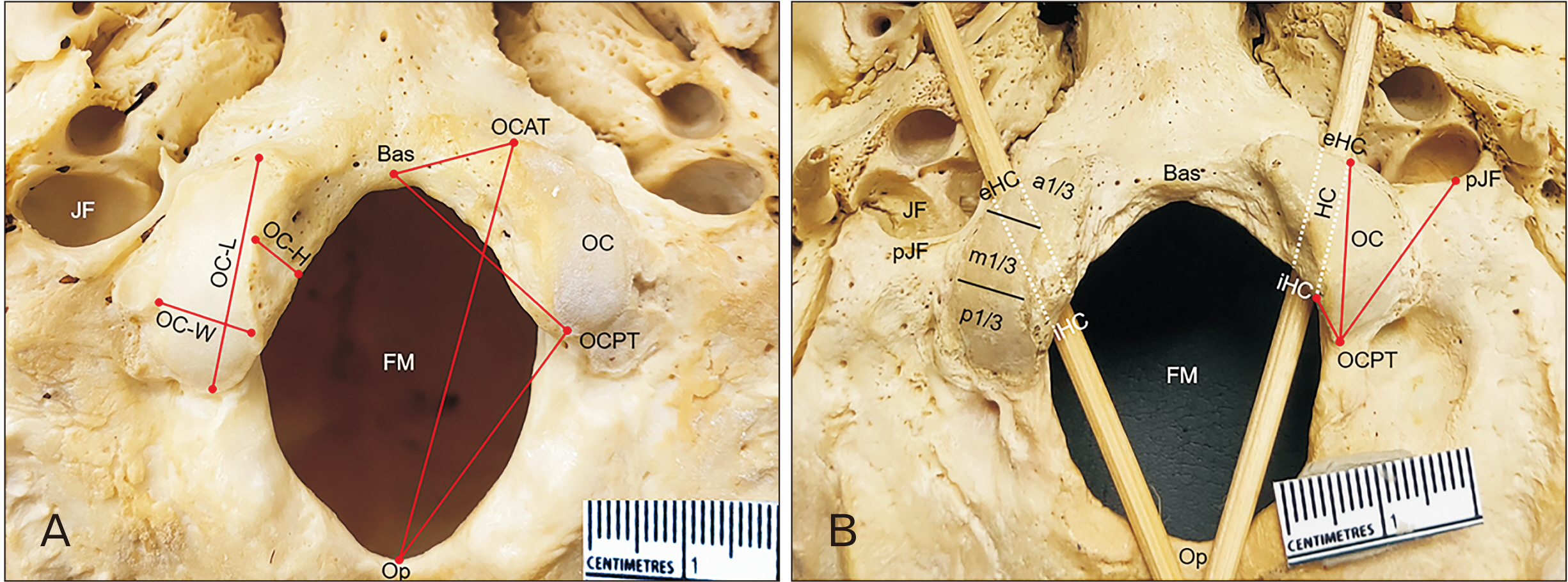
Fig. 1 Inferior view of the skull base. (A) Right occipital condyle showing occipital condyle width (OC-W), occipital condyle length (OC-L), and occipital condyle height (OC-H). Left occipital condyle showing the distances from anterior and posterior tips of occipital condyle (OCAT and OCPT) to basion (Bas) and opisthion (Op). (B) Right occipital condyle showing the relationship of jugular foramen (JF) and occipital condyle, the location of extracranial orifice and intracranial orifice of hypoglossal canal (L-eHC, L-iHC) related to occipital condyle. Left occipital condyle showing the distances from posterior tip of occipital condyle (OCPT) to iHC, eHC and posterior most end of jugular foramen (pJF). FM, foramen magnum; HC, hypoglossal canal; a1/3, anterior one third; m1/3, middle one third; p1/3, posterior one third.
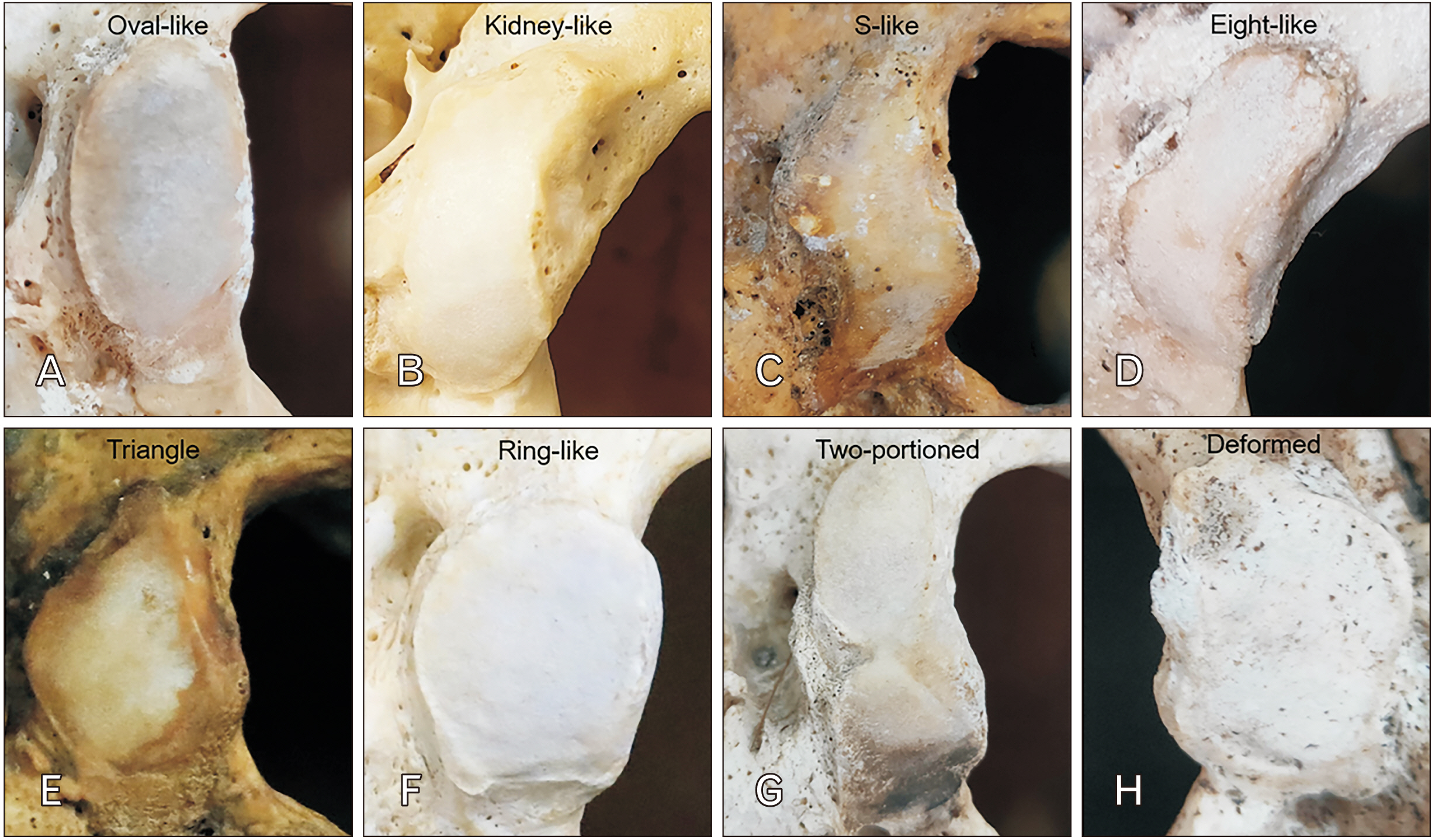
Fig. 2 Photographs showing eight shapes of occipital condyle. (A) Type I, oval-like condyle. (B) Type II, kidney-like condyle. (C) Type III, S-like condyle. (D) Type IV, eight-like condyle. (E) Type V, triangle condyle. (F) Type VI, ring-like condyle. (G) Type VII, two-portioned condyle, and (H) Type VIII, deformed condyle.
Cited by 1 articles
-
Morphological analysis of the jugular foramen in dry human skulls in northeastern Brazil
Rodrigo Ramalho Rodrigues, Diógenes Firmino do Nascimento Neto, João Vítor Andrade Fernandes, Letícia de Oliveira Barreto, Victor Barros Maciel do Amaral, Débora Karoline de Araújo Deca, Vera Louise Freire de Albuquerque Figueiredo, Jalles Dantas de Lucena, Ivson Bezerra da Silva, Thales Henrique de Araújo Sales, André de Sá Braga Oliveira
Anat Cell Biol. 2024;57(2):213-220. doi: 10.5115/acb.23.218.
Reference
-
References
1. Di G, Fang X, Hu Q, Zhou W, Jiang X. 2019; A microanatomical study of the far lateral approach. World Neurosurg. 127:e932–42. DOI: 10.1016/j.wneu.2019.04.004. PMID: 30995558.
Article2. Ayoub B. 2011; The far lateral approach for intra-dural anteriorly situated tumours at the craniovertebral junction. Turk Neurosurg. 21:494–8. DOI: 10.5137/1019-5149.JTN.4277-11.2. PMID: 22194106.
Article3. Rosa S, Baird JW, Harshfield D, Chehrenama M. 2018. Craniocervical junction syndrome: anatomy of the craniocervical and atlantoaxial junctions and the effect of misalignment on cerebrospinal fluid flow [Internet]. IntechOpen;London: Available from: https://www.intechopen.com/chapters/58783. cited 2022 Oct 18. DOI: 10.5772/intechopen.72890.4. lhan P, Kayhan B, Erturk M, Sengul G. 2017; Morphological analysis of occipital condyles and foramen magnum as a guide for lateral surgical approaches. MOJ Anat Physiol. 3:188–94. DOI: 10.15406/mojap.2017.03.00117.5. Mehdi W, Niaz A, Irfan M, Tasdique S, Majeed S. 2020; Far lateral transcondylar approach for anterior foramen magnum lesions. Pak J Neurol Surg. 24:149–55. DOI: 10.36552/pjns.v24i2.454.
Article6. Wu A, Zabramski JM, Jittapiromsak P, Wallace RC, Spetzler RF, Preul MC. 2010; Quantitative analysis of variants of the far-lateral approach: condylar fossa and transcondylar exposures. Neurosurgery. 66(6 Suppl Operative):191–8. discussion 198DOI: 10.1227/01.NEU.0000369704.49958.5B. PMID: 20489505.7. George JR, Francis T, Francis J, Samuel JE. 2019; Morphometric study of dry human occipital bone and its clinical relevance. Int J Anat Res. 7:6230–3. DOI: 10.16965/ijar.2018.447.
Article8. Scoville JP, Mazur MD, Couldwell WT. 2020; Unique far-lateral closure technique: technical note. Oper Neurosurg (Hagerstown). 18:384–90. DOI: 10.1093/ons/opz168. PMID: 31236599.
Article9. Moscovici S, Umansky F, Spektor S. 2015; "Lazy" far-lateral approach to the anterior foramen magnum and lower clivus. Neurosurg Focus. 38:E14. DOI: 10.3171/2015.2.FOCUS14784. PMID: 25828490.
Article10. Lanzino G, Paolini S, Spetzler RF. 2005; Far-lateral approach to the craniocervical junction. Neurosurgery. 57(4 Suppl):367–71. discussion 367–71. DOI: 10.1227/01.NEU.0000176848.05925.80. PMID: 16234687.
Article11. Chaddad-Neto F, Doria-Netto HL, Campos Filho JM, Reghin-Neto M, Rothon AL Jr. Oliveira Ed. 2014; The far-lateral craniotomy: tips and tricks. Arq Neuropsiquiatr. 72:699–705. DOI: 10.1590/0004-282X20140130. PMID: 25252234.
Article12. Cardoso AC, Fontes RB, Tan LA, Rhoton AL Jr, Roh SW, Fessler RG. 2015; Biomechanical effects of the transcondylar approach on the craniovertebral junction. Clin Anat. 28:683–9. DOI: 10.1002/ca.22551. PMID: 25914225.
Article13. Aragão JA, De Santana GM, Aragão ICS, Aragão FMS, Reis PF. Da Cruz de Moraes RZ. 2017; Morphological analysis on the occipital condyles and review of the literature. Int J Morphol. 35:1129–32. DOI: 10.4067/S0717-95022017000300050.
Article14. Anjum A, Pandurangam G, Garapati S, Bandarupalli N, Rabbani H, Divya P. 2021; Morphology and morphometric study of occipital condyles. Int J Anat Res. 9:7905–11. DOI: 10.16965/ijar.2021.107.
Article15. Karam YR, Traynelis VC. 2010; Occipital condyle fractures. Neurosurgery. 66(3 Suppl):56–9. DOI: 10.1227/01.NEU.0000365751.84075.66. PMID: 20173528. PMCID: PMC8571366.
Article16. Mazur MD, Couldwell WT, Cutler A, Shah LM, Brodke DS, Bachus K, Dailey AT. 2017; Occipitocervical instability after far-lateral transcondylar surgery: a biomechanical analysis. Neurosurgery. 80:140–5. DOI: 10.1093/neuros/nyw002. PMID: 28362894.
Article17. Lyrtzis C, Piagkou M, Gkioka A, Anastasopoulos N, Apostolidis S, Natsis K. 2017; Foramen magnum, occipital condyles and hypoglossal canals morphometry: anatomical study with clinical implications. Folia Morphol (Warsz). 76:446–57. DOI: 10.5603/FM.a2017.0002. PMID: 28150268.
Article18. Cheruiyot I, Mwachaka P, Saidi H. 2018; Morphometry of occipital condyles: implications for transcondylar approach to craniovertebral junction lesions. Anat J Afr. 7:1224–31. DOI: 10.4314/aja.v7i2.174142.
Article19. Verma R, Kumar S, Rai AM, Mansoor I, Mehra RD. 2016; The anatomical perspective of human occipital condyle in relation to the hypoglossal canal, condylar canal, and jugular foramen and its surgical significance. J Craniovertebr Junction Spine. 7:243–9. DOI: 10.4103/0974-8237.193258. PMID: 27891034. PMCID: PMC5111326.
Article20. Naderi S, Korman E, Citak G, Güvençer M, Arman C, Senoğlu M, Tetik S, Arda MN. 2005; Morphometric analysis of human occipital condyle. Clin Neurol Neurosurg. 107:191–9. DOI: 10.1016/j.clineuro.2004.07.014. PMID: 15823674.
Article21. Kaur J, ivastava D Sr, Singh D, Raheja S. 2012; The study of hyperostosic variants: significance of hyperostotic variants of human skulls in anthropology. Anat Cell Biol. 45:268–73. DOI: 10.5115/acb.2012.45.4.268. PMID: 23301194. PMCID: PMC3531590.
Article22. Ekanem UI, Olewnik Ł, Porzionato A, Macchi V, Iwanaga J, Loukas M, Dumont AS, Caro R, Tubbs RS. 2022; Morphology of the groove of the inferior petrosal sinus: application to better understanding variations and surgery of the skull base. Anat Cell Biol. 55:135–41. DOI: 10.5115/acb.22.023. PMID: 35773216. PMCID: PMC9256480.
Article23. Nagare SP, Chaudhari RS, Birangane RS, Parkarwar PC. 2018; Sex determination in forensic identification, a review. J Forensic Dent Sci. 10:61–6. DOI: 10.4103/jfo.jfds_55_17. PMID: 30745778. PMCID: PMC6344795.
Article24. Sangvichien S, Boonkaew K, Chuncharunee A, Komoltri C, Udom C, Chandee T. 2008; Accuracy of cranial and mandible morphological traits for sex determination in Thais. Siriraj Med J. 60:240–3. PMID: 09f90e8725f64b8eae26fd5fe5942626.25. Gilroy AM, MacPherson BR, Ross LM. 2008. Atlas of anatomy. 3rd ed. Thieme;Stuttgart: p. 742.26. Kalthur SG, Padmashali S, Gupta C, Dsouza AS. 2014; Anatomic study of the occipital condyle and its surgical implications in transcondylar approach. J Craniovertebr Junction Spine. 5:71–7. DOI: 10.4103/0974-8237.139201. PMID: 25210336. PMCID: PMC4158634.
Article27. Ozer MA, Celik S, Govsa F, Ulusoy MO. 2011; Anatomical determination of a safe entry point for occipital condyle screw using three-dimensional landmarks. Eur Spine J. 20:1510–7. DOI: 10.1007/s00586-011-1765-y. PMID: 21416278. PMCID: PMC3175895.
Article28. Bayat P, Bagheri M, Ghanbari A, Raoofi A. 2014; Characterization of occipital condyle and comparison of its dimensions with head and foramen magnum circumferences in dry skulls of Iran. Int J Morphol. 32:444–8. DOI: 10.4067/S0717-95022014000200011.
Article29. Saluja S, Das SS, Vasudeva N. 2016; Morphometric analysis of the occipital condyle and its surgical importance. J Clin Diagn Res. 10:AC01–4. DOI: 10.7860/JCDR/2016/23278.8800. PMID: 28050351. PMCID: PMC5198304. PMID: 02cb0daeaf6b4696acf783d82f99feef.30. Rai H, Keluskar V, Patil S, Bagewadi A. 2017; Accuracy of measurements of foramen magnum and occipital condyle as an indicator for sex determination using computed tomography. Indian J Health Sci Biomed Res. 10:80–3. DOI: 10.4103/2349-5006.198595.
Article31. Kumar A, Nagar M. 2014; Human adult occipital condyles: a morphometric analysis. Res Rev J Med Health Sci. 3:112–6.32. Vishteh AG, Crawford NR, Melton MS, Spetzler RF, Sonntag VK, Dickman CA. 1999; Stability of the craniovertebral junction after unilateral occipital condyle resection: a biomechanical study. J Neurosurg. 90(1 Suppl):91–8. DOI: 10.3171/spi.1999.90.1.0091. PMID: 10413132.
Article33. Shin H, Barrenechea IJ, Lesser J, Sen C, Perin NI. 2006; Occipitocervical fusion after resection of craniovertebral junction tumors. J Neurosurg Spine. 4:137–44. DOI: 10.3171/spi.2006.4.2.137. PMID: 16506481.
Article34. Karasu A, Cansever T, Batay F, Sabanci PA, Al-Mefty O. 2009; The microsurgical anatomy of the hypoglossal canal. Surg Radiol Anat. 31:363–7. DOI: 10.1007/s00276-008-0455-x. PMID: 19148566.
Article35. Bejjani GK, Sekhar LN, Riedel CJ. 2000; Occipitocervical fusion following the extreme lateral transcondylar approach. Surg Neurol. 54:109–15. discussion 115–6. DOI: 10.1016/S0090-3019(00)00255-X. PMID: 11077092.
Article36. Kizilkanat ED, Boyan N, Soames R, Oguz O. 2006; Morphometry of the hypoglossal canal, occipital condyle, and foramen magnum. Neurosurg Q. 16:121–5. DOI: 10.1097/01.wnq.0000214018.49915.49.
Article37. Parvindokht B, Reza DM, Saeid B. 2015; Morphometric analysis of hypoglossal canal of the occipital bone in Iranian dry skulls. J Craniovertebr Junction Spine. 6:111–4. DOI: 10.4103/0974-8237.161591. PMID: 26288545. PMCID: PMC4530509.
Article
- Full Text Links
-
- Actions
-
Cited
- CITED
-
- Close
- Share
-
- Similar articles
-
- Ventral Foramen Magnum Meningioma: Case Report
- Clinical and anatomical importance of foramen magnum and craniocervical junction structures in the perspective of surgical approaches
- Foramen Magnum Tumors
- A Case of Large Foramen Magnum Schwannoma
- Non-Metrical Morphologic Variations of Korean Skull Foramina
