Giant Cystic Cerebral Cavernous Malformation with Multiple Calcification: Case Report
- Affiliations
-
- 1Department of Neurosurgery, Cheongju St. Mary's hospital, Cheongju, Korea. k810a@hanmail.net
- KMID: 1491449
- DOI: http://doi.org/10.7461/jcen.2013.15.3.255
Abstract
- Cerebral cavernous malformation with giant cysts is rare and literature descriptions of its clinical features are few. In this case study, the authors describe the clinical symptoms, radiological findings, and pathological diagnosis of cerebral cavernous malformations with giant cysts, reviewing the relevant literature to clearly differentiate this from other disease entities. The authors present a case of a 19-year-old male with a giant cystic cavernous malformation, who was referred to the division of neurosurgery due to right sided motor weakness (grade II/II). Imaging revealed a large homogenous cystic mass, 7.2x4.6x6 cm in size, in the left fronto-parietal lobe and basal ganglia. The mass had an intra-cystic lesion, abutting the basal portion of the mass. The initial diagnosis considered this mass a glioma or infection. A left frontal craniotomy was performed, followed by a transcortical approach to resect the mass. Total removal was accomplished without post-operative complications. An open biopsy and a histopathological exam diagnosed the mass as a giant cystic cavernous malformation. Imaging appearances of giant cavernous malformations may vary. The clinical features, radiological features, and management of giant cavernous malformations are described based on pertinent literature review.
Keyword
MeSH Terms
Figure
-

Fig. 1 Preoperative imaging (A) Non-enhanced computed tomography scan shows a homogenous large-cystic lesion of 7.2×4.6×6 cm size in the left fronto-parietal lobe and basal ganglia. Note that there are mutliple parenchymal calcifications in both parietal lobes. (B) T1-weighted axial magnetic resonance imaging shows a large fronto-parietal hyperintense cystic lesion with local mass effect and minimal surrounding edema. (C) T2-weighted axial magnetic resonance imaging shows a heterogeneous high intra-cystic nodule, exhibited with hypodensities, suggestive of calcification. (D) T1-weighted contrast-enhanced coronal image reveals heterogeneous, slight enhancement of intra-cystic nodule, and a lack of enhancement of the cystic component.
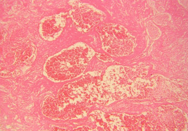
Fig. 2 Low-power photomicrographs show thromboses within the cavernous vascular spaces. Also note thin-walled vascular channels without neural tissue (Hematoxylin & Eosin, ×100).

Fig. 3 Postoperative computed tomography image shows no residual cavernous malformations with intact multiple calcifications.
Reference
-
1. Anderson RC, Connolly ES Jr, Ozduman K, Laurans MS, Gunel M, Khandji A, et al. Clinicopathological review: Giant intraventricular cavernous malformation. Neurosurgery. 2003; 8. 53(2):374–378. discussion 378-9. PMID: 12925254.
Article2. Avci E, Ozturk A, Baba F, Karabag H, Cakir A. Huge cavernoma with massive intracerebral hemorrhage in a child. Turk Neurosurg. 2007; 17(1):23–26. PMID: 17918674.3. Chicani CF, Miller NR, Tamargo RJ. Giant cavernous malformation of the occipital lobe. J Neuroophthalmol. 2003; 6. 23(2):151–153. PMID: 12782930.
Article4. Del Curling O Jr, Kelly DL Jr, Elster AD, Craven TE. An analysis of the natural history of cavernous angiomas. J Neurosurg. 1991; 11. 75(5):702–708. PMID: 1919691.
Article5. Duszak RS. Congenital rubella syndrome-Major review. Optometry. 2009; 1. 80(1):36–43. PMID: 19111256.
Article6. Engelhard HH, Stelea A, Mundt A. Oligodendroglioma and anaplastic oligodendroglioma: Clinical features, treatment, and prognosis. Surg Neurol. 2003; 11. 60(5):443–456. PMID: 14572971.7. Jones JL, Lopez A, Wilson M, Schulkin J, Gibbs R. Congenital toxoplasmosis: A review. Obstet Gynecol Surv. 2001; 5. 56(5):296–305. PMID: 11333376.
Article8. Kan P, Tubay M, Osborn A, Blaser S, Couldwell WT. Radiographic features of tumefactive giant cavernous angiomas. Acta Neurochir (Wien). 2008; 1. 150(1):49–55. discussion 55. PMID: 18066488.
Article9. Kenneson A, Cannon MJ. Review and meta-analysis of the epidemiology of congenital cytomegalovirus (CMV) infection. Rev Med Virol. 2007; Jul-Aug. 17(4):253–276. PMID: 17579921.
Article10. Kesava PP, Turski PA. MR angiography of vascular malformations. Neuroimaging Clin N Am. 1998; 5. 8(2):349–370. PMID: 9562593.11. Khosla VK, Banerjee AK, Mathuriya SN, Mehta S. Giant cystic cavernoma in a child. Case report. J Neurosurg. 1984; 6. 60(6):1297–1299. PMID: 6726375.12. Kim DS, Park YG, Choi JU, Chung SS, Lee KC. An analysis of the natural history of cavernous malformations. Surg Neurol. 1997; 7. 48(1):9–17. discussion 17-8. PMID: 9199678.
Article13. Lawton MT, Vates GE, Quinones-Hinojosa A, McDonald WC, Marchuk DA, Young WL. Giant infiltrative cavernous malformation: Clinical presentation, intervention, and genetic analysis: Case report. Neurosurgery. 2004; 10. 55(4):979–980. PMID: 15934180.
Article14. Martinez HR, Rangel-Guerra R, Elizondo G, Gonzalez J, Todd LE, Ancer J, et al. MR imaging in neuro-cysticercosis: A study of 56 cases. AJNR Am J Neuroradiol. 1989; Sep-Oct. 10(5):1011–1019. PMID: 2505513.15. Ohba S, Shimizu K, Shibao S, Nakagawa T, Murakami H. Cystic cavernous angiomas. Neurosurg Rev. 2010; 10. 33(4):395–400. PMID: 20174956.
Article16. Ramina R, Ingunza W, Vonofakos D. Cystic cerebral cavernous angioma with dense calcification. Case report. J Neurosurg. 1980; 2. 52(2):259–262. PMID: 7351568.17. Riant F, Bergametti F, Ayrignac X, Boulday G, Tournier-Lasserve E. Recent insights into cerebral cavernous malformations: The molecular genetics of CCM. FEBS J. 2010; 3. 277(5):1070–1075. PMID: 20096038.
Article18. Rigamonti D, Drayer BP, Johnson PC, Hadley MN, Zabramski J, Spetzler RF. The MRI appearance of cavernous malformations (angiomas). J Neurosurg. 1987; 10. 67(4):518–524. PMID: 3655889.
Article19. Sato K, Kubota T. Large calcified cystic cavernous angioma in the thalamus - Case report. Neurol Med Chir (Tokyo). 1995; 2. 35(2):100–103. PMID: 7753307.20. Simard JM, Garcia-Bengochea F, Ballinger WE Jr, Mickle JP, Quisling RG. Cavernous angioma: A review of 126 collected and 12 new clinical cases. Neurosurgery. 1986; 2. 18(2):162–172. PMID: 3960293.
Article21. van der Knaap MS, Vermeulen G, Barkhof F, Hart AA, Loeber JG, Weel JF. Pattern of white matter abnormalities at MR imaging: Use of polymerase chain reaction testing of Guthrie cards to link pattern with congenital cytomegalovirus infection. Radiology. 2004; 2. 230(2):529–536. PMID: 14752192.
Article
- Full Text Links
-
- Actions
-
Cited
- CITED
-
- Close
- Share
-
- Similar articles
-
- A Case of Cystic Cavernous Hemangioma
- A Case of Orbital Cavernous Angioma Associated with Intracranial Venous Anomalies
- A case involving anesthesia for cesarean section followed by resection of ruptured cavernous malformation of pons :A case report
- A Giant Aneurysmal Cerebral Arteriovenous Malformation in Childhood: Case Report
- Ruptured Cerebral Arteriovenous Malformation with Giant Venous Aneurysm: Case Report
