J Bacteriol Virol.
2008 Sep;38(3):119-125. 10.4167/jbv.2008.38.3.119.
Development of Capture ELISA Using a Biotinylated Monoclonal Antibody for Detection of Botulinum Neurotoxin Type A
- Affiliations
-
- 1Center for Infectious Diseases, National Institute of Health, 5-Nokbun-dong, Eunpyung-gu, Seoul, Korea. hboh@nih.go.kr
- KMID: 1483970
- DOI: http://doi.org/10.4167/jbv.2008.38.3.119
Abstract
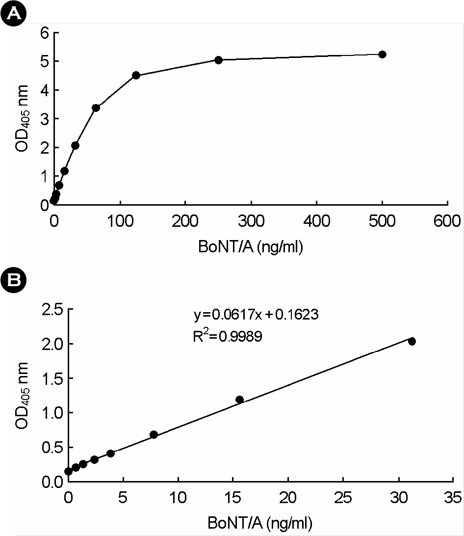
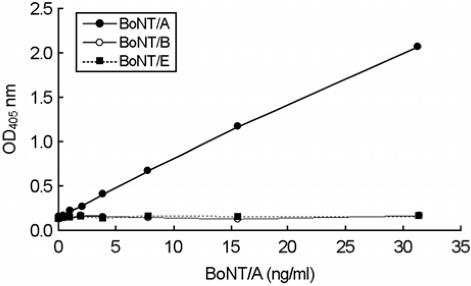
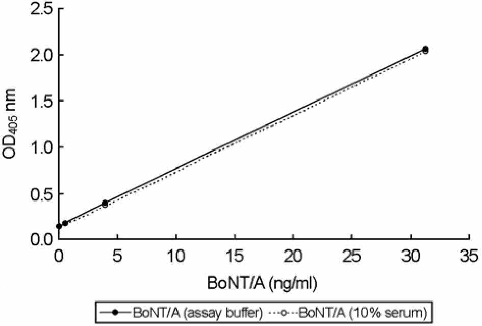
- A capture enzyme-linked immunosorbent assay (capture ELISA) was developed to detect Clostridium botulinum neurotoxin type A (BoNT/A) in assay buffer and human serum. The assay is based upon affinity-purified rabbit polyclonal and biotinylated monoclonal antibodies directed against the BoNT/A complex purified from C. botulinum ATCC19397. For the capture ELISA, the optimized amount (2 microgram/ml) of rabbit polyclonal antibody was immobilized on ELISA plates to detect BoNT/A (ranging from 0 to 500 ng/ml), which was recognized by 2 microgram/ml of the monoclonal antibody. From three independent repeated experiments, standard curves were linear over the range of 0~31.25 ng/ml BoNT/A and the coefficients (r(2)) ranged from 0.9951~0.9999 for all assays. The inter-variations were typically 0.50~6.93% and the specificity was confirmed by showing no cross-reactivity against BoNT/B and /E. The detection limit of capture ELISA was 0.488 ng/ml, which was close to mouse LD(50). In addition, application with BoNT/A-spiking human sera showed a possibility to detect BoNT/A with capture ELISA from the contaminated human sera. Taken together, the newly developed capture ELISA could serve as a rapid and sensitive screening tool for detecting BoNT/A simultaneously from massive specimens.
MeSH Terms
Figure
Reference
-
1). Boroff D., Fleck U. Statistical analysis of a rapid in vivo method for the titration of the toxin of Clostridium botulinum. J Bacteriol. 92:1580–1581. 1966.2). Cai S., Singh BR., Sharma S. Botulism diagnostics: from clinical symptoms to in vitro assays. Crit Rev Microbiol. 33:109–125. 2007.3). Center for Disease Control and prevention. Botulism in the United States, pp 1899-1996. Handbook for epidemiologists, clinicians, and laboratory workers. Center for Disease Control and Prevention, Atlanta GA. 1998.4). Chiao DJ., Wey JJ., Tang SS. Monoclonal antibody-based enzyme immunoassay for detection of botulinum neurotoxin type A. Hybridoma. 27:43–47. 2008.
Article5). Chung GT., Kang DH., Yoo CK., Choi JH., Seong WK. The first outbreak of botulism in Korea. Korean J Clin Microbiol. 6:160–163. 2003.6). Doellgast GJ., Triscott MX., Beard GA., Bottoms JD., Cheng T., Roh BH., Roman MG., Hall PA., Brown JE. Sensitive enzyme-linked immunosorbent assay for detection of Clostridium botulinum neurotoxins A, B, and E using signal amplification via enzyme-linked coagulation assay. J Clin Microbiol. 31:2402–2409. 1993.7). Geissler E., van Courtland Moon JE. Biological and toxin weapons: Research, development and use from the middle ages to 1945. Sipri Chemical & Biological Warfare Studies No. 18. Oxford University Press Inc;New York: 1999.8). Greenfield RA., Brown BR., Hutchins JB., Iandolo JJ., Jackson R., Slater LN., Bronze MS. Microbiological, biological, and chemical weapons of warfare and terrorism. Am J Med Sci. 323:326–340. 2002.
Article9). Heckels JE., Virji M. Separation and purification of surface components. pp. p. 67–135. In. Bacterial cell surface techniques. Hancock IC, Poxton IR, editors. (Ed),. John Wiley & Sons;Chichester, England: 1988.10). Lacy DB., Stevens RC. Sequence homology and structural analysis of the clostridial neurotoxins. J Mol Biol. 291:1091–1104. 1999.
Article11). Malizio CJ., Goodnough MC., Johnson EA. Purification of Clostridium botulinum type A neurotoxin. pp 27-39, Bacterial toxins: methods and protocols. Vol. 145:Holst O (Ed), Humana Press, Totowa, NJ,. 2000.12). Reed LJ., Muench H. A simple method of estimating fifty percent endpoints. Am J Hyg. 27:493–497. 1938.13). Sharma SK., Ferreira JL., Eblen BS., Whiting RC. Detection of type A, B, E, and F Clostridium botulinum neurotoxins in foods by using an amplified enzyme-linked immunosorbent assay with digoxigenin-labeled antibodies. Appl Environ Microbiol. 72:1231–1238. 2006.14). Shone C., Wilton-Smith P., Appleton N., Hambleton P., Modi N., Gatley S., Melling J. Monoclonal antibody-based immunoassay for type A Clostridium botulinum toxin is comparable to the mouse bioassay. Appl Environ Microbiol. 50:63–67. 1985.15). Simpson LL. Botulinum neurotoxin and tetanus toxin. Academic Press, San Diego. 1989.16). Szilagyi M., Rivera VR., Neal D., Merrill GA., Poli MA. Development of sensitive colorimetric capture elisas for Clostridium botulinum neurotoxin serotypes A and B. Toxicon. 38:381–389. 2000.17). Yi HA., Lim JG., Lee JB., Her JH., Kim HA., Shin YE., Cho YW., Lee H., Yi SD. A familial outbreak of food-borne botulism. J Korean Neurol Assoc. 22:670–672. 2004.
- Full Text Links
- Actions
-
Cited
- CITED
-
- Close
- Share
- Similar articles
-
- Development of Enzyme Linked Immunosorbent Assay for Erythropoietin
- Molecular Mechanism of the Action of Clostridium botulinum Type B Neurotoxin
- Detection of Platelet-Specific Antibodies Employing Modified Antigen Capture ELISA(MACE)
- Detection of Infectious Bursal Disease Virus by Double Antibody Sandwich ELISA
- Development of immunoassays for the detection of kanamycin in veterinary fields


