MRI-Guided Intervention for Breast Lesions Using the Freehand Technique in a 3.0-T Closed-Bore MRI Scanner: Feasibility and Initial Results
- Affiliations
-
- 1Department of Radiology, Gyeongsang National University Hospital, Jinju 660-702, Korea.
- 2Department of Radiology, Seoul National University Bundang Hospital, Seongnam 463-707, Korea. kimsmlms@daum.net
- 3Department of Surgery, Seoul National University Bundang Hospital, Seongnam 463-707, Korea.
- 4Department of Pathology, Seoul National University Bundang Hospital, Seongnam 463-707, Korea.
- 5Department of Radiology, Seoul National University College of Medicine, Seoul National University Hospital, Seoul 110-744, Korea.
- 6Department of Radiology, Samsung Medical Center, Seoul 135-710, Korea.
- KMID: 1482774
- DOI: http://doi.org/10.3348/kjr.2013.14.2.171
Abstract
OBJECTIVE
To report the feasibility of magnetic resonance imaging (MRI)-guided intervention for diagnosing suspicious breast lesions detectable by MRI only, using the freehand technique with a 3.0-T closed-bore MRI scanner.
MATERIALS AND METHODS
Five women with 5 consecutive MRI-only breast lesions underwent MRI-guided intervention: 3 underwent MRI-guided needle localization and 2, MRI-guided vacuum-assisted biopsy. The interventions were performed in a 3.0-T closed-bore MRI system using a dedicated phased-array breast coil with the patients in the prone position; the freehand technique was used. Technical success and histopathologic outcome were analyzed.
RESULTS
MRI showed that four lesions were masses (mean size, 11.5 mm; range, 7-18 mm); and 1, a nonmass-like enhancement (maximum diameter, 21 mm). The locations of the lesions with respect to the breast with index cancer were as follows: different quadrant, same breast - 3 cases; same quadrant, same breast - 1 case; and contralateral breast - 1 case. Histopathologic evaluation of the lesions treated with needle localization disclosed perilobular hemangioma, fibrocystic change, and fibroadenomatous change. The lesions treated with vacuum-assisted biopsy demonstrated a radial scar and atypical apocrine hyperplasia. Follow-up MRI after 2-7 months (mean, 4.6 months) confirmed complete lesion removal in all cases.
CONCLUSION
MRI-guided intervention for breast lesions using the freehand technique with a 3.0-T closed-bore MRI scanner is feasible and accurate for diagnosing MRI-only lesions.
Keyword
MeSH Terms
-
Adult
Biopsy, Needle
Breast Neoplasms/*pathology
Contrast Media/diagnostic use
Diagnosis, Differential
Feasibility Studies
Female
Gadolinium DTPA/diagnostic use
Humans
Magnetic Resonance Imaging/*instrumentation
Magnetic Resonance Imaging, Interventional/*methods
Middle Aged
Neoplasm Staging
Retrospective Studies
Vacuum
Contrast Media
Gadolinium DTPA
Figure
-

Fig. 1 Breast MRI equipment and technique. Patient was placed in prone position (A) on dedicated phased-array bilateral breast coil. Handheld MRI-guided biopsy device (arrows in B) was introduced into coaxial sheath (arrow in C) to acquire tissue specimens.
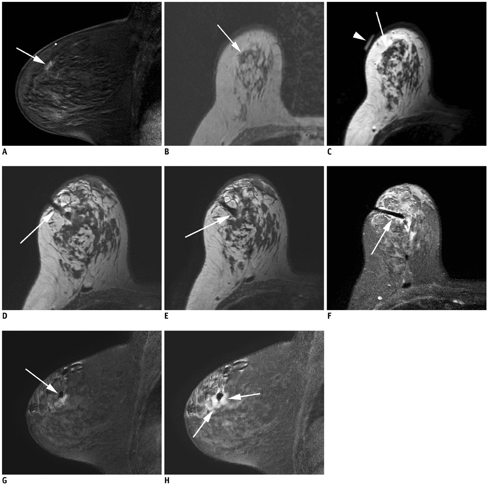
Fig. 2 60-year-old woman diagnosed with invasive ductal carcinoma in left breast after US-guided core needle biopsy. A, B. Preoperative MRI scan shows 1.0 cm ill-defined irregular early-enhancing mass (arrow) in right breast on sagittal post Gd-enhanced fat-suppressed T1-weighted image (A) and axial non-enhanced T1-weighted image, which was not seen in second-look US. To exclude bilateral breast cancer, MRI-guided vacuum-assisted biopsy was performed. C. Noncontrast axial T1-weighted image before needle placement showed lesion with low T1 signal intensity (arrow) under skin marker (arrowhead). D, E. Nonenhanced axial T1-weighted images following needle placement showed that needle tip (arrow) was advanced to edge of lesion, ideal position for accurate sampling. F, G. Axial and sagittal contrast-enhanced images confirmed correct needle placement (arrows). H. Post-biopsy, sagittal fat-suppressed T1-weighted image showed high signal intensity surrounding needle due to hematoma, air, and anesthetic (arrows). Pathologic examination of cores indicated atypical apocrine hyperplasia. This high-risk lesion was referred to surgery following US-guided needle localization for hematoma at site of previous biopsy. No residual lesion was found on pathologic examination. US = ultrasound
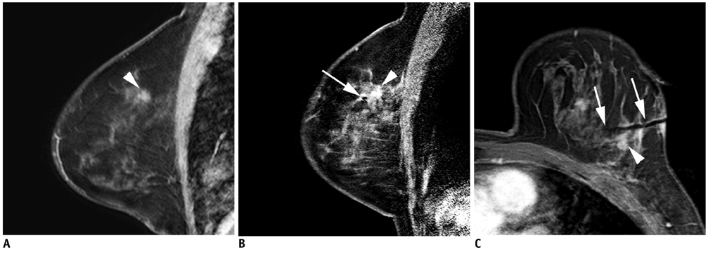
Fig. 3 56-year-old woman diagnosed with invasive ductal carcinoma in left breast after US-guided core needle biopsy. A. Sagittal T1-weighted contrast-enhanced MR image shows 11-mm ill-defined irregular early-enhancing mass (arrowhead) in different quadrant of same breast with index cancer. No corresponding lesion was detected on second-look US. To exclude multicentric growth, MRI-guided tissue sampling was performed by needle localization followed by surgical excision. B, C. Sagittal (B) and axial (C) contrast-enhanced images obtained after needle placement showed needle tip (arrow) just anterior to enhancing mass (arrowhead). Lesion was successfully localized and excisional biopsy showed fibroadenomatous changes. US = ultrasound
Cited by 1 articles
-
Initial Experience with Magnetic Resonance-Guided Vacuum-Assisted Biopsy in Korean Women with Breast Cancer
Hye Na Jung, Boo-Kyung Han, Eun Young Ko, Jung Hee Shin
J Breast Cancer. 2014;17(3):270-278. doi: 10.4048/jbc.2014.17.3.270.
Reference
-
1. Orel SG, Schnall MD, LiVolsi VA, Troupin RH. Suspicious breast lesions: MR imaging with radiologic-pathologic correlation. Radiology. 1994. 190:485–493.2. Stomper PC, Herman S, Klippenstein DL, Winston JS, Edge SB, Arredondo MA, et al. Suspect breast lesions: findings at dynamic gadolinium-enhanced MR imaging correlated with mammographic and pathologic features. Radiology. 1995. 197:387–395.3. Boné B, Aspelin P, Bronge L, Isberg B, Perbeck L, Veress B. Sensitivity and specificity of MR mammography with histopathological correlation in 250 breasts. Acta Radiol. 1996. 37:208–213.4. Daniel BL, Yen YF, Glover GH, Ikeda DM, Birdwell RL, Sawyer-Glover AM, et al. Breast disease: dynamic spiral MR imaging. Radiology. 1998. 209:499–509.5. Kuhl CK, Elevelt A, Leutner CC, Gieseke J, Pakos E, Schild HH. Interventional breast MR imaging: clinical use of a stereotactic localization and biopsy device. Radiology. 1997. 204:667–675.6. Daniel BL, Birdwell RL, Ikeda DM, Jeffrey SS, Black JW, Block WF, et al. Breast lesion localization: a freehand, interactive MR imaging-guided technique. Radiology. 1998. 207:455–463.7. Brenner RJ, Shellock FG, Rothman BJ, Giuliano A. Technical note: magnetic resonance imaging-guided pre-operative breast localization using "freehand technique". Br J Radiol. 1995. 68:1095–1098.8. Lee CH, Smith RC, Levine JA, Troiano RN, Tocino I. Clinical usefulness of MR imaging of the breast in the evaluation of the problematic mammogram. AJR Am J Roentgenol. 1999. 173:1323–1329.9. Morris EA, Liberman L, Dershaw DD, Kaplan JB, LaTrenta LR, Abramson AF, et al. Preoperative MR imaging-guided needle localization of breast lesions. AJR Am J Roentgenol. 2002. 178:1211–1220.10. Kuhl CK, Bieling H, Gieseke J, Ebel T, Mielcarek P, Far F, et al. Breast neoplasms: T2* susceptibility-contrast, first-pass perfusion MR imaging. Radiology. 1997. 202:87–89.11. Daniel BL, Birdwell RL, Butts K, Nowels KW, Ikeda DM, Heiss SG, et al. Freehand iMRI-guided large-gauge core needle biopsy: a new minimally invasive technique for diagnosis of enhancing breast lesions. J Magn Reson Imaging. 2001. 13:896–902.12. van den Bosch MA, Daniel BL, Pal S, Nowels KW, Birdwell RL, Jeffrey SS, et al. MRI-guided needle localization of suspicious breast lesions: results of a freehand technique. Eur Radiol. 2006. 16:1811–1817.13. Meeuwis C, Peters NH, Mali WP, Gallardo AM, van Hillegersberg R, Schipper ME, et al. Targeting difficult accessible breast lesions: MRI-guided needle localization using a freehand technique in a 3.0 T closed bore magnet. Eur J Radiol. 2007. 62:283–288.14. Hauth EA, Jaeger HJ, Lubnau J, Maderwald S, Otterbach F, Kimmig R, et al. MR-guided vacuum-assisted breast biopsy with a handheld biopsy system: clinical experience and results in postinterventional MR mammography after 24 h. Eur Radiol. 2008. 18:168–176.15. van de Ven SM, Lin MC, Daniel BL, Sareen P, Lipson JA, Pal S, et al. Freehand MRI-guided preoperative needle localization of breast lesions after MRI-guided vacuum-assisted core needle biopsy without marker placement. J Magn Reson Imaging. 2010. 32:101–109.16. American College of Radiology (ACR). BI-RADS® Breast Imaging Reporting and Data System, Breast Imaging Atlas. 2003. Reston, VA: American College of Radiology.17. Ikeda DM, Hylton NM, Kinkel K, Hochman MG, Kuhl CK, Kaiser WA, et al. Development, standardization, and testing of a lexicon for reporting contrast-enhanced breast magnetic resonance imaging studies. J Magn Reson Imaging. 2001. 13:889–895.18. Veltman J, Boetes C, Wobbes T, Blickman JG, Barentsz JO. Magnetic resonance-guided biopsies and localizations of the breast: initial experiences using an open breast coil and compatible intervention device. Invest Radiol. 2005. 40:379–384.19. Fischer U, Kopka L, Grabbe E. Magnetic resonance guided localization and biopsy of suspicious breast lesions. Top Magn Reson Imaging. 1998. 9:44–59.20. Pfleiderer SO, Reichenbach JR, Azhari T, Marx C, Wurdinger S, Kaiser WA. Dedicated double breast coil for magnetic resonance mammography imaging, biopsy, and preoperative localization. Invest Radiol. 2003. 38:1–8.21. Smith LF, Henry-Tillman R, Rubio IT, Korourian S, Klimberg VS. Intraoperative localization after stereotactic breast biopsy without a needle. Am J Surg. 2001. 182:584–589.22. Fischer U, Vosshenrich R, Keating D, Bruhn H, Döler W, Oestmann JW, et al. MR-guided biopsy of suspect breast lesions with a simple stereotaxic add-on-device for surface coils. Radiology. 1994. 192:272–273.23. Heywang-Köbrunner SH, Heinig A, Pickuth D, Alberich T, Spielmann RP. Interventional MRI of the breast: lesion localisation and biopsy. Eur Radiol. 2000. 10:36–45.24. Orel SG, Schnall MD, Newman RW, Powell CM, Torosian MH, Rosato EF. MR imaging-guided localization and biopsy of breast lesions: initial experience. Radiology. 1994. 193:97–102.25. Carbognin G, Girardi V, Brandalise A, Baglio I, Bucci A, Bonetti F, et al. MR-guided vacuum-assisted breast biopsy in the management of incidental enhancing lesions detected by breast MR imaging. Radiol Med. 2011. 116:876–885.26. Youk JH, Kim EK, Kim MJ, Ko KH, Kwak JY, Son EJ, et al. Concordant or discordant? Imaging-pathology correlation in a sonography-guided core needle biopsy of a breast lesion. Korean J Radiol. 2011. 12:232–240.27. Döler W, Fischer U, Metzger I, Harder D, Grabbe E. Stereotaxic add-on device for MR-guided biopsy of breast lesions. Radiology. 1996. 200:863–864.28. Coulthard A. Magnetic resonance imaging-guided preoperative breast localization using a "freehand technique". Br J Radiol. 1996. 69:482–483.29. Kuhl CK, Morakkabati N, Leutner CC, Schmiedel A, Wardelmann E, Schild HH. MR imaging--guided large-core (14-gauge) needle biopsy of small lesions visible at breast MR imaging alone. Radiology. 2001. 220:31–39.
- Full Text Links
-
- Actions
-
Cited
- CITED
-
- Close
- Share
-
- Similar articles
-
- MRI-Guided Breast Intervention: Biopsy and Needle Localization
- Clinical Applications of Breast MRI
- Breast Magnetic Resonance Imaging-Guided Biopsy
- The Role of Preoperative Breast MRI in Patients With Early-Stage Breast Cancer
- Tips for finding magnetic resonance imaging-detected suspicious breast lesions using second-look ultrasonography: a pictorial essay
