Tuberc Respir Dis.
2008 May;64(5):369-373. 10.4046/trd.2008.64.5.369.
A Case of Multiple Micronodular Pneumocyte Hyperplasia of the Lung in a Man with Tuberous Sclerosis
- Affiliations
-
- 1Department of Internal Medicine, National Health Insurance Corporation Ilsan Hospital, Goyang, Korea. hch7001@nhimc.or.kr
- 2Department of Pathology, National Health Insurance Corporation Ilsan Hospital, Goyang, Korea.
- 3Department of Radiology, National Health Insurance Corporation Ilsan Hospital, Goyang, Korea.
- KMID: 1478193
- DOI: http://doi.org/10.4046/trd.2008.64.5.369
Abstract
- Tuberous sclerosis (TS) is an autosomal dominant disorder that is characterized by cutaneous lesions, seizures, mental retardation and hamartomas in various organs including the skin, kidney and brain. Pulmonary involvement is extremely rare, and occurs in approximately 0.1 to 1% of TS cases. Recent reports have indicated multiple micronodular pneumocyte hyperplasia (MMPH) as another rare form of pulmonary involvement of tuberous sclerosis. We report a case of a 35 year-old-male patient who had no pulmonary symptoms but showed multinodular pulmonary shadows on his chest CT scan. The patient was finally diagnosed with TS with MMPH of the lung. MMPH does not appear to have any malignant potential but the clinical significance of MMPH in TS patients is unknown.(Tuberc Respir Dis 2008;64:369-373)
MeSH Terms
Figure
-
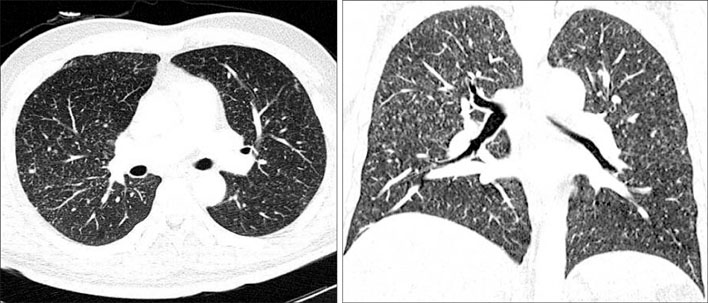
Figure 1 Chest CT scan shows randomly scattered multiple nodules throughout both lungs with upper lung predominance.
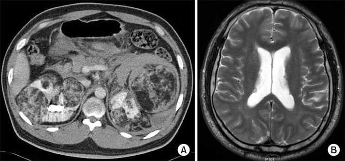
Figure 2 Abdominopelvic CT scan demonstrates variable sized multiple renal angiomyolipoma with focal rupture causing large amount of retroperitoneal and peritoneal hemorrhage (A). Brain MR image shows several subependymal nodules in the ventricular walls of the lateral ventricle on T2-weighted image (B).
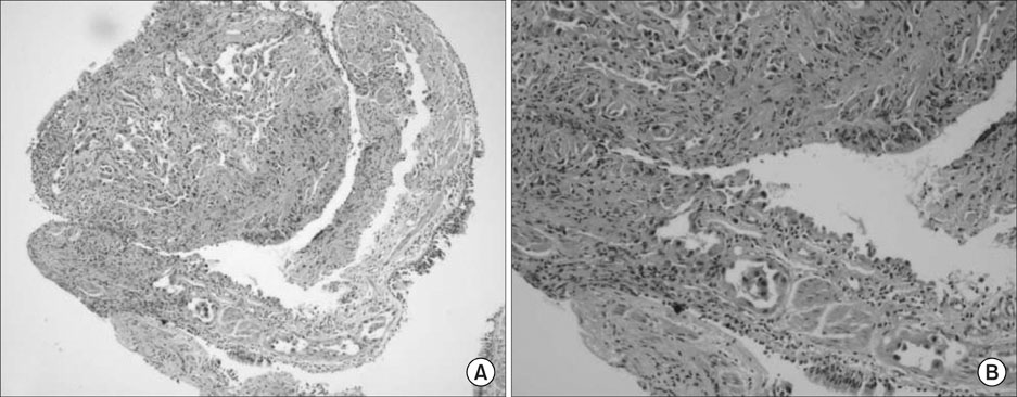
Figure 3 Microscopic view of the transbronchial lung biopsy specimen shows well demarcated multiple nodules of cuboidal type II pneumocytes along the fibrously thickened alveolar septa (H&E stain, A, ×100, B, ×400).
Reference
-
1. Sparagana SP, Roach ES. Tuberous sclerosis complex. Curr Opin Neurol. 2000. 13:115–119.2. Yagci C, Sahin-Akyar G, Akyar S. Multiple organ involvement in tuberous sclerosis. Eur J Radiol. 1997. 25:52–54.3. Costello LC, Hartman TE, Ryu JH. High frequency of pulmonary lymphangioleiomyomatosis in women with tuberous sclerosis complex. Mayo Clin Proc. 2000. 75:591–594.4. Maruyama H, Ohbayashi C, Hino O, Tsutsumi M, Konishi Y. Pathogenesis of multifocal micronodular pneumocyte hyperplasia and lymphangioleiomyomatosis in tuberous sclerosis and association with tuberous sclerosis genes TSC1 and TSC2. Pathol Int. 2001. 51:585–594.5. Maruyama H, Seyama K, Sobajima J, Kitamura K, Sobajima T, Fukuda T, et al. Multifocal micronodular pneumocyte hyperplasia and lymphangioleiomyomatosis in tuberous sclerosis with a TSC2 gene. Mod Pathol. 2001. 14:609–614.6. Popper HH, Juettner-Smolle FM, Pongratz MG. Micronodular hyperplasia of type II pneumocytes-a new lung lesion associated with tuberous sclerosis. Histopathology. 1991. 18:347–354.7. Mo EK, Jung MP, Yoo CG, Kim YW, Han SK, Im JG, et al. Lymphangioleiomyomatosis in Korea. Tuberc Respir Dis. 1993. 40:519–531.8. Baik JM, Hong HK, Oh YB, Lee SM, Park MS, Yoo TK, et al. A case of pulmonary lymphangioleiomyomatosis associated with tuberous sclerosis and renal angiomyolipoma. Tuberc Respir Dis. 1997. 44:1184–1193.9. Shen A, Iseman MD, Waldron JA, King TE. Exacerbation of pulmonary lymphangioleiomyomatosis by exogenous estrogens. Chest. 1987. 91:782–785.10. Brentani MM, Carvalho CR, Saldiva PH, Pacheco MM, Oshima CT. Steroid receptors in pulmonary lymphangiomyomatosis. Chest. 1984. 85:96–99.11. Moss J, Avila NA, Barnes PM, Litzenberger RA, Bechtle J, Brooks PG, et al. Prevalence and clinical characteristics of lymphangioleiomyomatosis (LAM) in patients with tuberous sclerosis complex. Am J Respir Crit Care Med. 2001. 164:669–671.12. Franz DN, Brody A, Meyer C, Leonard J, Chuck G, Dabora S, et al. Mutational and radiographic analysis of pulmonary disease consistent with lymphangioleiomyomatosis and micronodular pneumocyte hyperplasia in women with tuberous sclerosis. Am J Respir Crit Care Med. 2001. 164:661–668.13. Fujitaka K, Isobe T, Oguri T, Yamasaki M, Miyazaki M, Kohno N, et al. A case of micronodular pneumocyte hyperplasia diagnosed through lung biopsy using thoracoscopy. Respiration. 2002. 69:277–279.14. Bonetti F, Pea M, Martignoni G, Doglioni C, Zamboni G, Capelli P, et al. Clear cell ("sugar") tumor of the lung is a lesion strictly related to angiomyolipoma--the concept of a family of lesions characterized by the presence of the perivascular epithelioid cells (PEC). Pathology. 1994. 26:230–236.15. Fujitaka K, Isobe T, Oguri T, Yamasaki M, Miyazaki M, Kohno N, et al. A case of micronodular pneumocyte hyperplasia diagnosed through lung biopsy using thoracoscopy. Respiration. 2002. 69:277–279.
- Full Text Links
-
- Actions
-
Cited
- CITED
-
- Close
- Share
-
- Similar articles
-
- Rare Lung Manifestation of Multifocal Micronodular Pneumocyte Hyperplasia in a Teenage Girl with Tuberous Sclerosis Complex
- Pulmonary Lymphangioleiomyomatosis and Micronodular Pneumocyte Hyperplasia associated with Tuberous Sclerosis: A Case Report
- Concurrent renal and hepatic angiomyolipoma with pulmonary involvement in two patients with tuberous sclerosis
- A Case of Unilateral Renal Angiomyolipoma Associated with Tuberous Sclerosis
- Hamartomatous gastric polyposis in a patient with tuberous sclerosis
