Gossypiboma Encountered 40 Years after Lumbar Partial Laminectomy: A Case Report
- Affiliations
-
- 1Department of Orthopedic Surgery, Handong University Sunlin Hospital, Pohang, Korea.
- 2Department of Neurosurgery, Handong University Sunlin Hospital, Pohang, Korea.
- 3Department of Orthopedic Surgery, Wallace Memorial Baptist Hospital, Busan, Korea. msh124@paran.com
- KMID: 1473391
- DOI: http://doi.org/10.4184/jkss.2009.16.1.54
Abstract
- Gossypiboma is a mass within body consisting of a cotton matrix surrounded by a foreign-body reaction. Some patients may remain asymptomatic, while others develop early persistent infected conditions. Gossypiboma should be included in a differential diagnosis of a paravertebral mass in postoperative patients, and a thorough and a careful inspection of the surgical field before closure must be performed by surgeons to avoid the complications of gossypiboma even when there are correct counts. We present a patient in whom a gossypiboma at the 4th lumbar spine was encountered 40 years after a partial laminectomy with no subjective symptoms.
Keyword
Figure
-
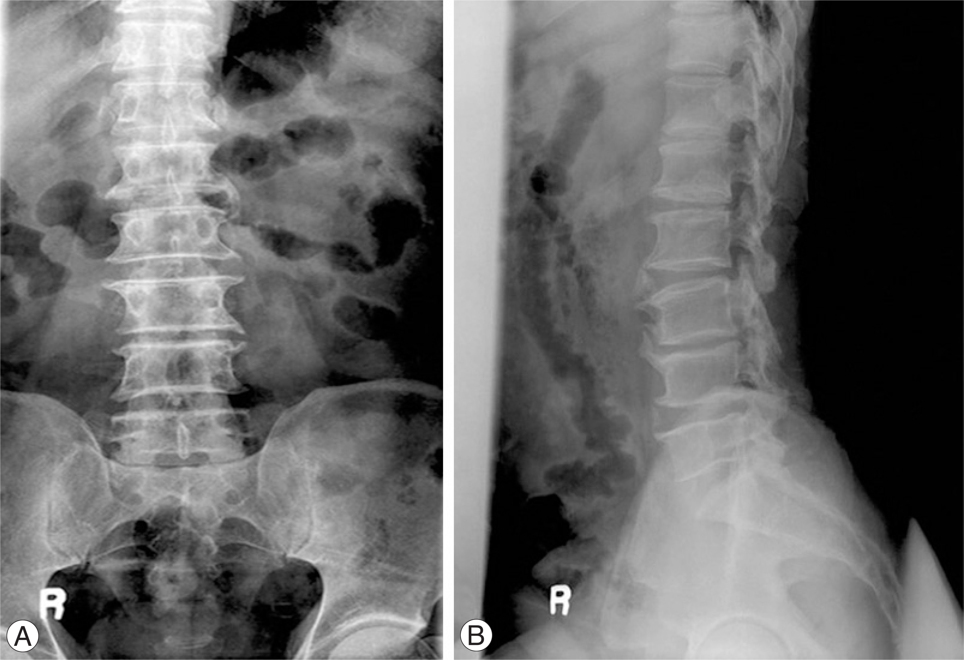
Fig. 1. A 60-year-old man who underwent left L3 and L4 partial laminectomy 40 years ago. Preoperative (A) anteroposterior and (B) lateral radiographs show left laminectomy state of L3 and L4.

Fig. 2. (A) Sagittal and (B) axial T1 weighted images show paravertebral mass with an intermediate signal intensity (arrows). (C) Sagittal and (D) axial T2 weighted images show a high signal intensity within the center of the lesion and a low signal intensity within the peripheral rim (arrows). (E) Sagittal and (F) axial enhanced images reveal strong enhancement of the peripheral rim of the lesion (arrows).
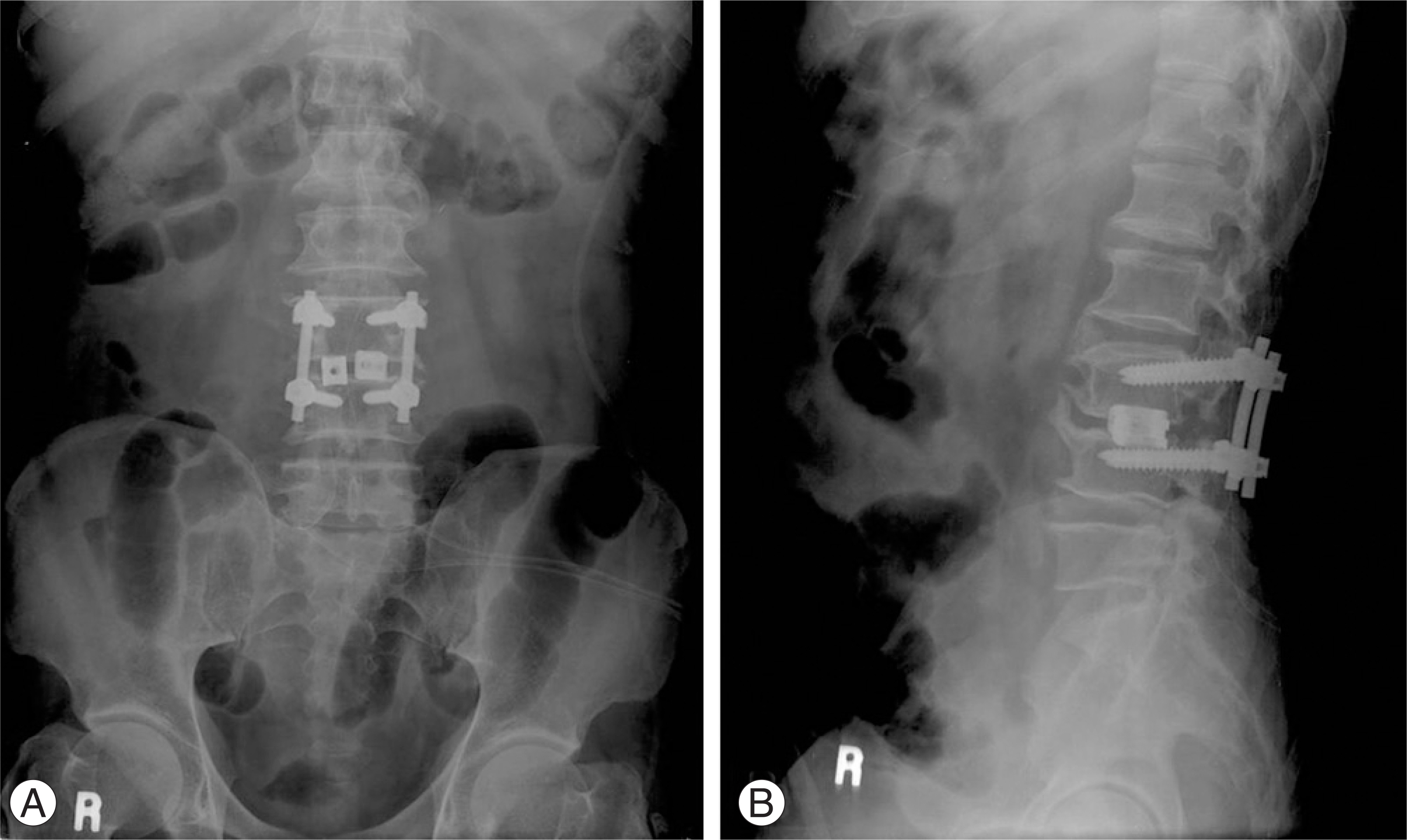
Fig. 3. Postoperative (A) anteroposterior and (B) lateral radiographs show posterior lumbar intervertebral fusion and posterolateral fusion state of L3 and L4.
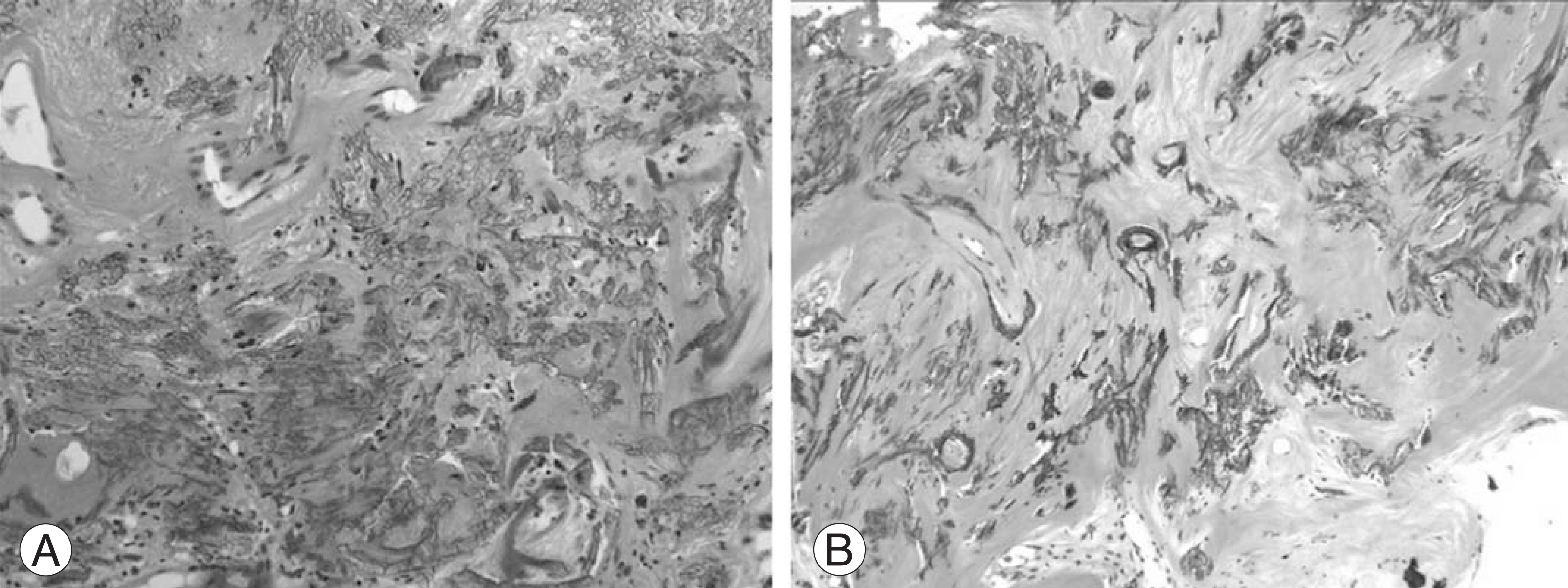
Fig. 4. (A) Foreign materials area scattered in the sclerotic stroma with mild infiltration of lymphocytes. (B) Multiple microcalcifications are present (Hematoxylin & eosin stain, × 200).
Reference
-
01). Rajput A., Loud PA., Gibbs JF., Kraybill WG. Diagnostic challenges in patients with tumors: case 1. Gossypiboma (foreign body) manifesting 30 years after laparotomy. J Clin Oncol. 2003. 21:3700–3701.02). Suh DH., Kim EC. Pathologic fracture of femoral neck due to mass suspicious of gossypiboma in proximal thigh. -case report-. J Korean Hip Soc. 2006. 18:493–497.
Article03). Rappaport W., Haynes K. The retained surgical sponge following intra-abdominal surgery. A continuing problem. Arch Surg. 1990. 125:405–407.04). Bevernage C., Geusens E., Nijs S. Case report: a gossypiboma in the shoulder. Emerg Radiol. 2006. 12:231–233.
Article05). IS M., Karatas A., Akgul M., Yildirim U., Gezen F. A retained surgical sponge (gossypiboma) mimicking a paraspinal abscess. Br J Neurosurg. 2007. 21:307–308.
Article06). Van Goethem JW., Parizel PM., Perdius D., Hermans P., De Moor J. MR and CT imaging of paraspinal textilo-ma(gossypiboma). J Comput Assist Tomogr. 1991. 15:1000–1003.07). Yuh-Feng T., Chin-Chu W., Cheng-Tau S., Min-Tsung T. FDG PET CT features of an intraabdominal gossypiboma. Clin Nucl Med. 2005. 30:561–563.
Article
- Full Text Links
-
- Actions
-
Cited
- CITED
-
- Close
- Share
-
- Similar articles
-
- Intracranial Gossypiboma Mimicking a Recurrent Low Grade Astrocytoma: Case Report
- Pathologic Fracture of Femoral Neck due to Mass suspicious of Gossypiboma in Proximal Thigh: Case Report
- Postlaminectomy Bilateral Lumbar Intraspinal Synovial Cysts
- Retroperitoneal Gossypiboma
- A Case Report of Neglected Gossypiboma Causing Abdominal Pain for 20 Years Post-Cesarean Section
