Efficiency of Anterior Interbody Fusion using Cage Packed with DBM in the Distractive Flexion Injury of Cervical Spine: Demineralized Bone Matrix vs Autoiliac Cancellous Bone
- Affiliations
-
- 1Department of Orthopaedic Surgery, Yonsei University, Wonju College of Medicine, Wonju, Korea. par73@wonju.yonsei.ac.kr
- KMID: 1459276
- DOI: http://doi.org/10.4184/jkss.2010.17.3.111
Abstract
- STUDY DESIGN: This is a retrospective study.
OBJECTIVES
We wanted to evaluate the effectiveness and safety of a PEEK cage filled with DBM in patients with a distractive flexion injury of the cervical spine. SUMMARY OF LITERATURE REVIEW: AIF of the cervical spine using an autoiliac bone graft and plate fixation is known to be an effective treatment for traumatic injuries. However, the complications arising from the donor site are troublesome, and so fusion with cage is an alternative treatment.
MATERIALS AND METHODS
We analyzed 32 cases (22 males and 10 females) with distractive flexion injury of the cervical spine. They underwent anterior decompression and interbody fusion with a PEEK cage and anterior plate fixation. In 18 patients, the cage was filled with autogenous iliac bone (Group I), and for the other 10 the cages were filled with DBM (Group II).
RESULTS
All the cases in Group I and Group II achieved fusion except for one case of nonunion in group II. The anterior and posterior vertebral heights of the fused segments of group II were decreased more than those of group I, resulting a statistical difference (p=0.003). The changes of segmental lordosis (p=0.69) and the neurologic status (p=0.22) showed no statistical difference between the two groups.
CONCLUSIONS
AIF using a PEEK cage filled with DBM and plate fixation showed no significant clinical differences compared to the case of iliac bone autografting. However, from a radiologic perspective, the time to achieve union was extensive and a case of nonunion was also observed. Therefore, many considerations are necessary when using DBM as a replacement for iliac bone autografting and further research should be done on this subject.
MeSH Terms
Figure
-
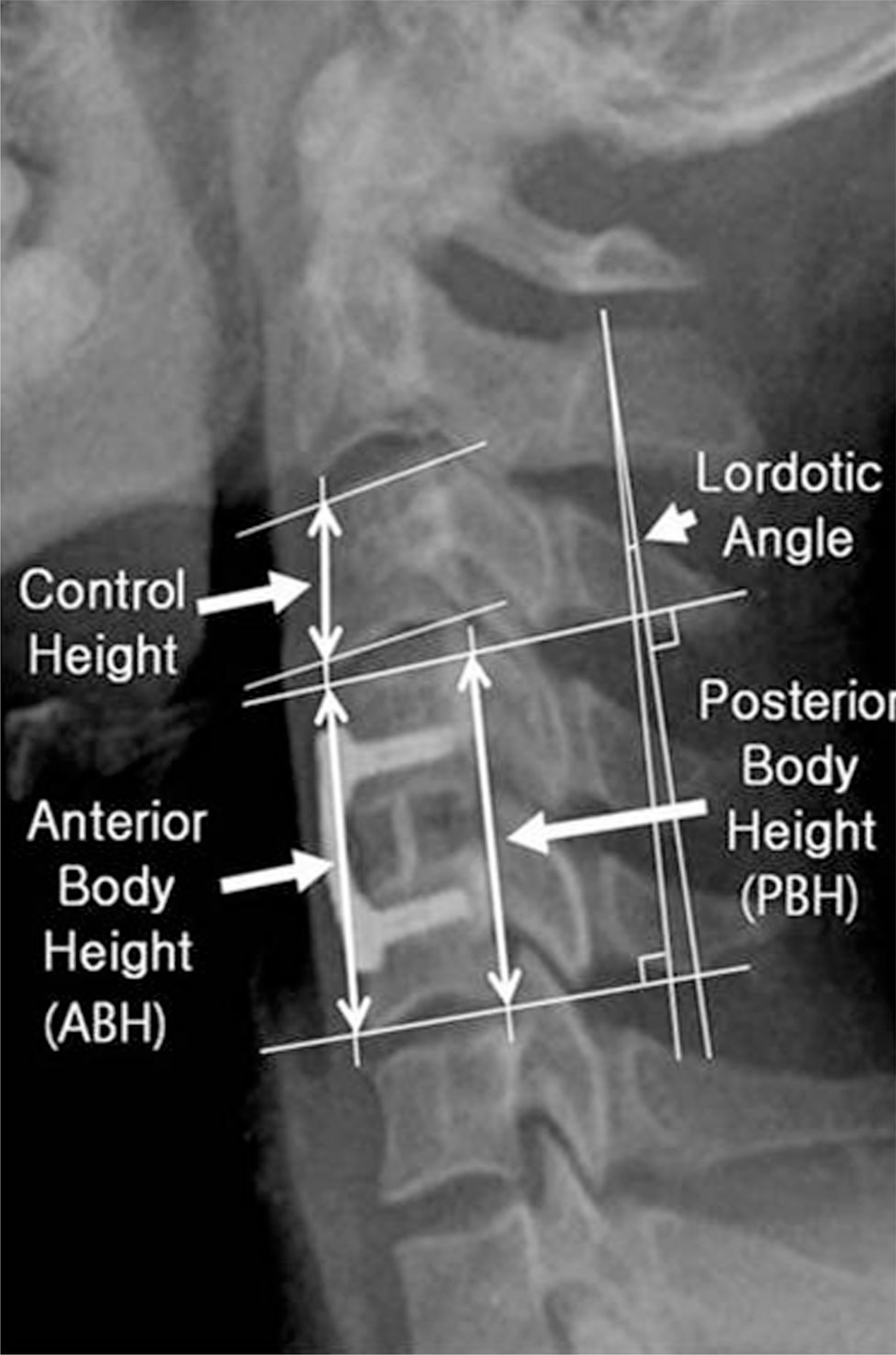
Fig. 1. Radiograph showing linear and angular measurement.
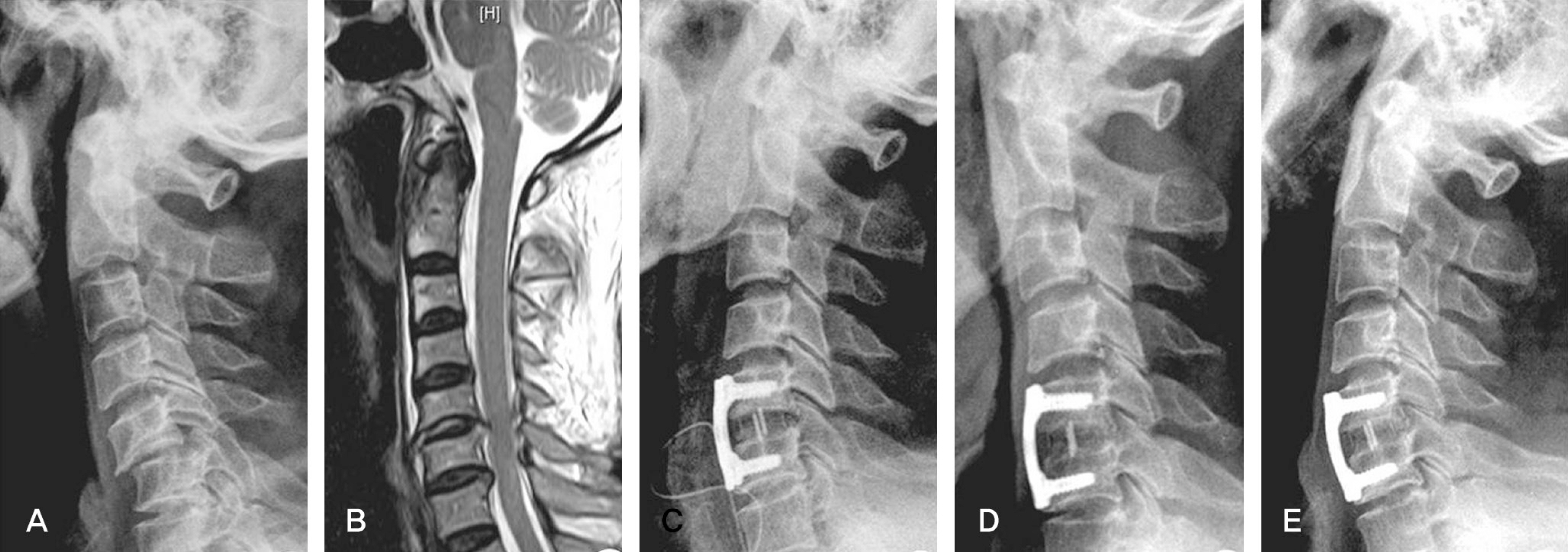
Fig. 2. A 43-year old male with C5 nerve root injury due to bilateral facet dislocation. (A) Preoperative lateral roentgenogram shows anterior displacement of C5 on C6 body. (B) T2 weighted sagittal MR image shows a C5-6 disc protrusion. (C) Lateral radiograph, immediately after surgery, shows anterior cervical fusion with cervical spine locking plate and Solis PEEK cage that packed with cancellous iliac bone. (D,E) Lateral roentgenogram of flexion/extension views that show no motion and solid fusion.

Fig. 3. A 19-year old male with C5 nerve root injury due to unilateral facet dislocation. (A) Preoperative lateral roentgenogram shows anterior displacement of C4 on C5 body. (B) T2 weighted sagittal MR image shows a C4-5 disc protrusion. (C) Lateral radiograph, immediately after surgery, shows anterior cervical fusion with cervical spine locking plate and Solis PEEK cage that packed with demineralized bone matrix. (D,E) Lateral roentgenogram of flexion/extension views that show no motion and solid fusion.
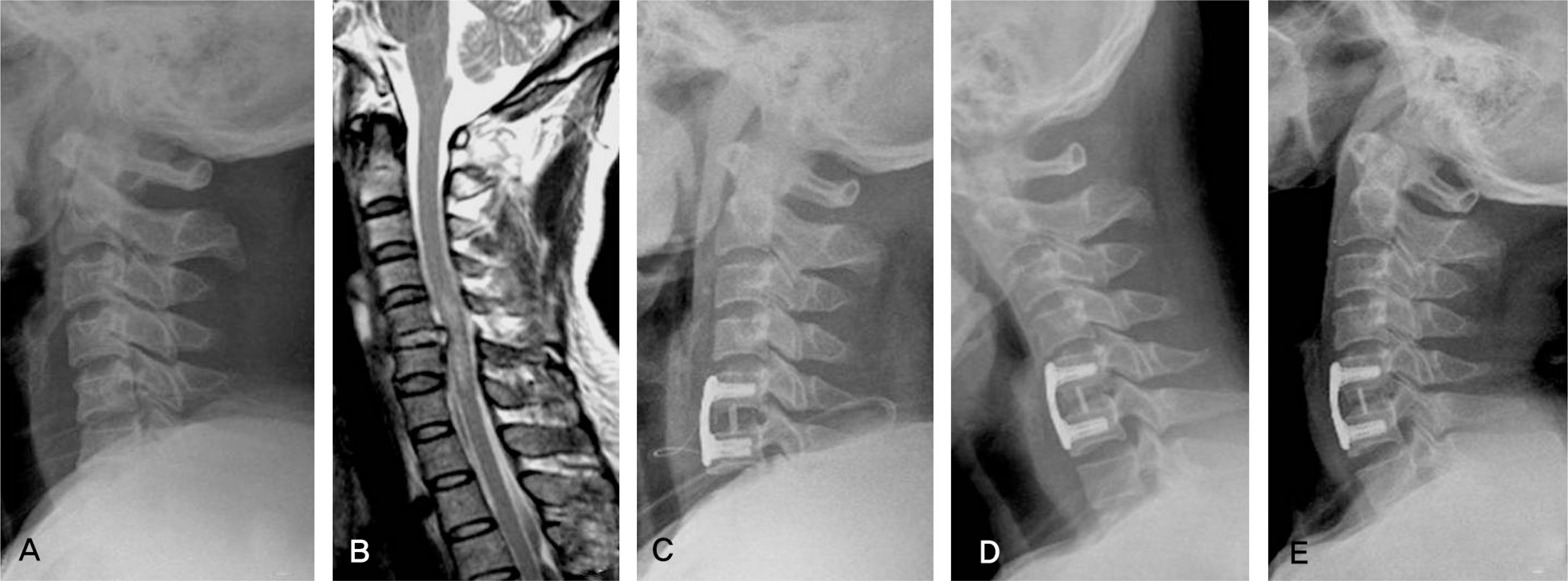
Fig. 4. A 49-year old female with spinal cord injury due to unilateral facet dislocation. (A) Preoperative lateral roentgenogram shows anterior displacement of C5 on C6 body. (B) T2 weighted sagittal MR image shows a C5-6 disc protrusion and a change of spinal cord signal. (C) Lateral radiograph, immediately after surgery, shows anterior cervical fusion with cervical spine locking plate and Solis PEEK cage that packed with demineralized bone matrix. (D,E) Lateral roentgenogram of flexion/extension views that show transverse radiolucent line and 5.2˚ fusion segmental motion.
Reference
-
1.Vaccaro AR., Stubbs HA., Block JE. Demineralized bone matrix composite grafting for posterolateral spinal fusion. Orthopedics. 2007. 30:567–70.2.An HS., Simpson JM., Glover JM., Stephany J. Comparison between allograft plus demineralized bone matrix versus autograft in anterior cervical fusion. A prospective multicenter study. Spine. 1995. 20:2211–16.3.Sassard WR., Eidman DK., Gray PM, et al. Augmenting local bone with Grafton demineralized bone matrix for posterolateral lumbar spine fusion: avoiding second site autologous bone harvest. Orthopedics. 2000. 23:1059–64.
Article4.Girardi FP., Cammisa FP Jr. The effect of bone graft extenders to enhance the performance of iliac crest bone grafts in instrumented lumbar spine fusion. Orthopedics. 2003. 26:S545–8.
Article5.Louis-Ugbo J., Murakami H., Kim HS., Minamide A., Boden SD. Evidence of osteoinduction by Grafton demineralized bone matrix in nonhuman primate spinal fusion. Spine. 2004. 29:360–6.
Article6.Choi DJ., Ahn DK., Lee S, et al. The effect of demineralized bone matrix as a graft enhancer in posterior lumbar interbody fusion using cage and local bone chips. J Korean Soc Spine Surg. 2008. 15:157–64.
Article7.Habal MB., Reddi AH. Bone grafts and bone induction substitutes. Clin Plast Surg. 1994. 21:525–42.
Article8.Malloy KM., Hilibrand AS. Autograft versus allograft in degenerative cervical disease. Clin Orthop Relat Res. 2002. 394:27–38.
Article9.Arrington ED., Smith WJ., Chambers HG., Bucknell AL., Davino NA. Complications of iliac crest bone graft harvesting. Clin Orthop Relat Res. 1996. 329:300–9.
Article10.Silber JS., Anderson DG., Daffner SD, et al. Donor site morbidity after anterior iliac crest bone harvest for single-level anterior cervical discectomy and fusion. Spine. 2003. 28:134–9.
Article11.Summers BN., Eisenstein SM. Donor site pain from the ilium. A complication of lumbar spine fusion. J Bone Joint Surg Br. 1989. 71:677–80.
Article12.Rawlinson JN. Morbidity after anterior cervical decompression and fusion. The influence of the donor site on recovery, and the results of a trial of surgibone compared to autologous bone. Acta Neurochir (Wien). 1994. 131:106–18.
Article13.Cho DY., Lee WY., Sheu PC. Treatment of multilevel cervical fusion with cages. Surg Neurol. 2004. 62:378–85.
Article14.Zdeblick TA., Ducker TB. The use of freeze-dried allograft bone for anterior cervical fusions. Spine. 1991. 16:726–9.
Article15.Deutsch H., Haid R., Rodts G Jr., Mummaneni PV. The decision-making process: allograft versus autograft. Neurosurgery. 2007. 60:S98–102.16.Bibbo C., Patel DV. The effect of demineralized bone matrix-calcium sulfate with vancomycin on calcaneal fracture healing and infection rates: a prospective study. Foot Ankle Int. 2006. 27:487–93.
Article17.Urban RM., Turner TM., Hall DJ., Infanger S., Cheema N., Lim TH. Healing of large defects treated with calcium sulfate pellets containing demineralized bone matrix particles. Orthopedics. 2003. 26:S581–5.
Article18.Turner TM., Urban RM., Hall DJ., Cheema N., Lim TH. Restoration of large bone defects using a hard-setting, injectable putty containing demineralized bone particles compared to cancellous autograft bone. Orthopedics. 2003. 26:S561–5.
Article19.Galjour C., Dzugan S., Graves M, et al. Stimulation of fracture healing by continuous delivery of demineralized bone matrix proteins and tobramycin. Biomed Sci Instrum. 2005. 41:122–7.20.Martin GJ Jr., Boden SD., Titus L., Scarborough NL. New formulations of demineralized bone matrix as a more effective graft alternative in experimental posterolateral lumbar spine arthrodesis. Spine. 1999. 24:637–45.
Article21.Russell JL., Block JE. Clinical utility of demineralized bone matrix for osseous defects, arthrodesis, and reconstruction: impact of processing techniques and study methodology. Orthopedics. 1999. 22:524–31.22.Ijiri S., Yamamuro T., Nakamura T., Kotani S., Notoya K. Effect of sterilization on bone morphogenetic protein. J Orthop Res. 1994. 12:628–36.
Article23.Drosos GI., Kazakos KI., Kouzoumpasis P., Verettas DA. Safety and efficacy of commercially available demineralised bone matrix preparations: a critical review of clinical studies. Injury. 2007. 38:S13–21.
Article24.Edwards JT., Diegmann MH., Scarborough NL. Osteoinduction of human demineralized bone: character-ization in a rat model. Clin Orthop Relat Res. 1998. 357:219–228.25.Frederic S., Benedict R., Payer M. Implantation of an empty carbon fiber cage or a tricortical iliac crest autograft after cervical discectomy for single-level disc herniation: a prospective comparative study. J Neurosurg Spine. 2006. 4:292–9.26.Payer M., May D., Reverdin A., Tessitore E. Implantation of an empty carbon fiber composite frame cage after single-level anterior cervical discectomy in the treatment of cervical disc herniation: preliminary results. J Neurosurg. 2003. 98:143–8.
Article27.Salame K., Ouaknine GE., Razon N., Rochkind S. The use of carbon fiber cages in anterior cervical interbody fusion: report of 100 cases. Neurosurg Focus. 2002. 12:E1.28.Schroder J., Schul C., Hasselblatt M., Wassmann H. Bony fusion through an empty cervical disc interspace implant. Zentralbl Neurochir. 2007. 68:139–41.29.Park HJ., Kim WK., Ryu HY. Efficiency of anterior interbody fusion using cage and plate in the distractive flexion injury of cervical spine: cage vs tricortical autoiliac bone. J Korean Soc Spine Surg. 2009. 16:71–8.
- Full Text Links
-
- Actions
-
Cited
- CITED
-
- Close
- Share
-
- Similar articles
-
- Efficiency of Anterior Interbody Fusion using Cage and Plate in the Distractive Flexion Injury of Cervical Spine : Cage vs Tricortical Autoiliac Bone
- Outcomes of Anterior Cervical Fusion Using Polyetheretherketone Cage with Demineralized Bone Matrix and Plate for Management of Subaxial Cervical Spine Injuries
- The Effect of Demineralized Bone Matrix as a Graft Enhancer in Posterior Lumbar Interbody Fusion Using Cage and Local Bone Chips
- Surgical Outcomes of Anterior Cervical Fusion Using Deminaralized Bone Matrix as Stand-Alone Graft Material: Single Arm, Pilot Study
- Polyetheretherketone Cage with Demineralized Bone Matrix Can Replace Iliac Crest Autografts for Anterior Cervical Discectomy and Fusion in Subaxial Cervical Spine Injuries
