Central odontogenic fibroma: a case report
- Affiliations
-
- 1Department of Oral and Maxillofacial Radiology, School of Dentistry, Pusan National University, Busan, Korea. ksnah@pusan.ac.kr
- KMID: 1449940
- DOI: http://doi.org/10.5624/isd.2011.41.2.85
Abstract
- Central odontogenic fibroma is a rare odontogenic neoplasm that originates from odontogenic ectomesenchyme. Here, a case of central odontogenic fibroma in a 17-year-old male is reported. Since the present case showed a multilocular radiolucency with partially ill-defined border between the right mandibular condyle and the distal root of the right mandibular third molar, differential diagnosis involved a wide range of pathosis from benign lesions like ameoloblastic fibroma and odontogenic myxoma to more aggressive lesions such as desmoplastic fibroma, juvenile aggressive fibromatosis, or fibrosarcoma.
Keyword
MeSH Terms
Figure
-
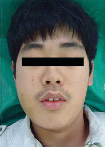
Fig. 1 Extraoral photograph shows a swelling on the right cheek causing facial asymmetry.

Fig. 2 Intraoral photograph shows normal-colored mucosal right cheek swelling extending to the retromolar area.

Fig. 3 Cropped panoramic image shows a multilocular radiolucency with partially ill-defined border between the right mandibular condyle and the distal root of the right mandibular third molar.

Fig. 4 Axial CT image shows an expansile solid ovoid mass involving the right mandibular condyle and ramus measuring around 3.5×3.2 cm.

Fig. 5 Axial CT image shows slight enhancement of the lesion.
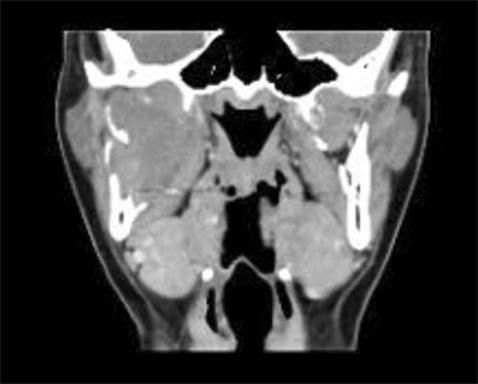
Fig. 6 Coronal CT image shows massive bone resorption of the right mandibular condylar process and ramus.
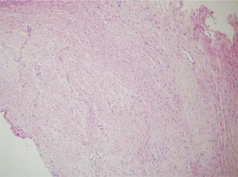
Fig. 7 Photomicrograph reveals a non-encapsulated tumor consisting of bland fibroblast cells. A few strands and nests of odontogenic epithelium is observed (H&E stain, ×100).
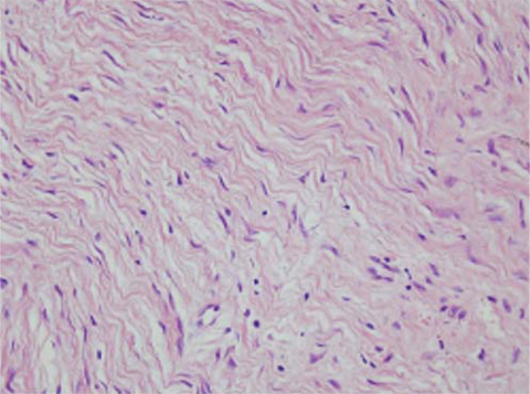
Fig. 8 Photomicrograph reveals bland fibroblast cells with wavy cytoplasm (H&E stain, ×400).
Cited by 1 articles
-
Central odontogenic fibroma (simple type) in a four-year-old boy: atypical cone-beam computed tomographic appearance with periosteal reaction
Najme Anbiaee, Hamed Ebrahimnejad, Alireza Sanaei
Imaging Sci Dent. 2015;45(2):109-115. doi: 10.5624/isd.2015.45.2.109.
Reference
-
1. Neville BW, Damm DD, Allen CM, Bouquot JE. Oral and maxillofacial pathology. 2002. 2nd ed. Philadelphia: WB Saunders;633–635.2. Regezi JA, Kerr DA, Courtney RM. Odontogenic tumors; analysis of 706 cases. J Oral Surg. 1978. 36:771–778.3. Handlers JP, Abrams AM, Melrose RJ, Danforth R. Central odontogenic fibroma: clinicopathologic features of 19 cases and review of the literature. J Oral Maxillofac Surg. 1991. 49:46–54.
Article4. Daley TD, Wysocki GP, Pringle GA. Relative incidence of odontogenic tumors and oral and jaw cysts in a Canadian population. Oral Surg Oral Med Oral Pathol. 1994. 77:276–280.
Article5. Buchner A, Merrell PW, Carpenter WM. Relative frequency of central odontogenic tumors: a study of 1,088 cases from Northern California and comparison to studies from other parts of the world. J Oral Maxillofac Surg. 2006. 64:1343–1352.
Article6. Lin HP, Chen HM, Vu CH, Yang H, Kuo RC, Kuo YS, et al. Odontogenic fibroma: a clinicopathological study of 15 cases. J Formos Med Assoc. 2011. 110:27–35.
Article7. Buchner A, Merrell PW, Carpenter WM. Relative frequency of peripheral odontogenic tumors: a study of 45 new cases and comparison with studies from the literature. J Oral Pathol Med. 2006. 35:385–391.
Article8. Covani U, Crespi R, Perrini N, Barone A. Central odontogenic fibroma: a case report. Med Oral Patol Oral Cir Bucal. 2005. 10:Suppl 2. E154–E157.9. Philipsen HP, Reichart PA, Sciubba JJ, van der Waal I. Barnes L, Eveson JW, Reichart PA, Sidransky D, editors. Odontogenic fibroma. World Health Organization classification of tumours. Pathology and genetics of tumours of head and neck tumours. 2005. Lyon: IARC;317.10. Daniels JS. Central odontogenic fibroma of mandible: a case report and review of the literature. Oral Surg Oral Med Oral Pathol Oral Radiol Endod. 2004. 98:295–300.
Article11. Gardner DG. Central odontogenic fibroma current concepts. J Oral Pathol Med. 1996. 25:556–561.
Article12. Daskala I, Kalyvas D, Kolokoudias M, Vlachodimitropoulos D, Alexandridis C. Central odontogenic fibroma of the mandible: a case report. J Oral Sci. 2009. 51:457–461.
Article13. Kaffe I, Buchner A. Radiologic features of central odontogenic fibroma. Oral Surg Oral Med Oral Pathol. 1994. 78:811–818.
Article14. Araki M, Nishimura S, Matsumoto N, Ohnishi M, Ohki H, Komiyama K. Central odontogenic fibroma with osteoid formation showing atypical radiographic appearance. Dentomaxillofac Radiol. 2009. 38:426–430.
Article
- Full Text Links
-
- Actions
-
Cited
- CITED
-
- Close
- Share
-
- Similar articles
-
- Central odontogenic fibroma of the simple type
- Aggressive central odontogenic fibroma in the maxilla: A case report
- Central Odontogenic Fibroma in Anterior Maxilla: A Case Report
- Central odontogenic fibroma case report
- Central odontogenic fibroma (simple type) in a four-year-old boy: atypical cone-beam computed tomographic appearance with periosteal reaction
