Central odontogenic fibroma (simple type) in a four-year-old boy: atypical cone-beam computed tomographic appearance with periosteal reaction
- Affiliations
-
- 1Department of Oral and Maxillofacial Radiology, Maxillofacial Diseases Research Center, School of Dentistry, Mashhad University of Medical Sciences, Mashhad, Iran. ebrahimnh911@mums.ac.ir
- KMID: 2054213
- DOI: http://doi.org/10.5624/isd.2015.45.2.109
Abstract
- Central odontogenic fibroma (COF) is a rare benign tumor that accounts for 0.1% of all odontogenic tumors. A case of COF (simple type) of the mandible in a four-year-old boy is described in this report. The patient showed asymptomatic swelling in the right inferior border of the lower jaw for one week. A panoramic radiograph showed a poorly-defined destructive unilocular radiolucent area. Cone-beam computed tomography showed expansion and perforation of the adjacent cortical bone plates. A periosteal reaction with the Codman triangle pattern was clearly visible in the buccal cortex. Since the tumor had destroyed a considerable amount of bone, surgical resection was performed. No recurrence was noted.
MeSH Terms
Figure
-

Fig. 1 A panoramic radiograph shows a poorly-defined radiolucent lesion in the posterior mandibular region, perforating the inferior mandibular cortex.
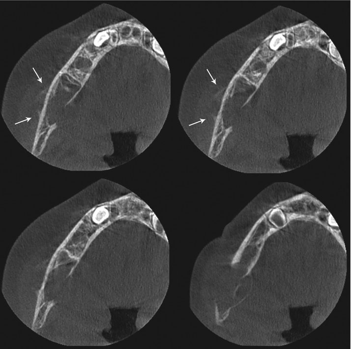
Fig. 2 Axial cone-beam computed tomography images show lingual expansion and perforation of the buccal and lingual cortical plates. Note the periosteal reaction (Codman triangle) in the buccal cortex (arrows).

Fig. 3 Panoramic (A) and cross-sectional (B) reformatted cone-beam computed tomography images show a multilocular lesion with some faint septae. The lesion has perforated the lingual, buccal, and inferior mandibular cortical plates, the cortical outlines of the follicles of the molars, and the inferior alveolar nerve canal. Periosteal new bone formation is seen in the buccal cortical plate (arrows).
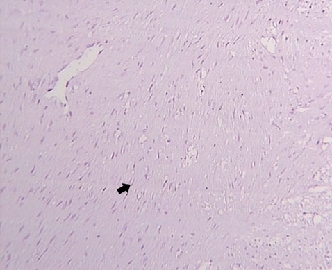
Fig. 4 Plump fibroblasts (arrow) within a collagenous background. No epithelial remnants are found (H&E stain, 100×)

Fig. 5 Fibrous stroma with fibroblastic cells. Some myxoid changes (asterisks) in the stroma, normal bone trabecules containing osteocyte lacunae (arrowheads), and osteoblastic margins (arrows) are seen (H&E stain, 100×)

Fig. 6 Postoperative panoramic radiograph.

Fig. 7 One-month follow-up radiograph. The infected bone graft had been removed.
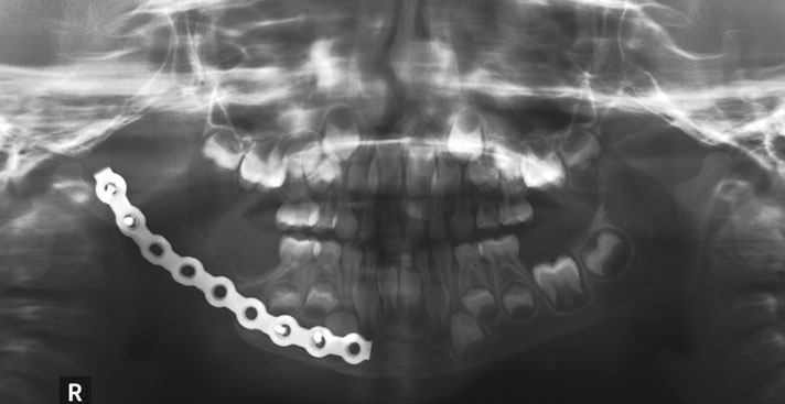
Fig. 8 A six-month follow-up panoramic radiograph shows the patient's stable condition with no recurrence.
Reference
-
1. Daniels JS. Central odontogenic fibroma of mandible: a case report and review of the literature. Oral Surg Oral Med Oral Pathol Oral Radiol Endod. 2004; 98:295–300.
Article2. Kaffe I, Buchner A. Radiologic features of central odontogenic fibroma. Oral Surg Oral Med Oral Pathol. 1994; 78:811–818.
Article3. Covani U, Crespi R, Perrini N, Barone A. Central odontogenic fibroma: a case report. Med Oral Patol Oral Cir Bucal. 2005; 10:Suppl 2. E154–E157.4. Cicconetti A, Bartoli A, Tallarico M, Maggiani F, Santaniello S. Central odontogenic fibroma interesting the maxillary sinus. A case report and literature survey. Minerva Stomatol. 2006; 55:229–239.5. Barnes L, Eveson JW, Reichart P, Sidransky D. World Health Organization classification of tumours. Pathology and genetics: head and neck tumours. Lyon: IARC Press;2005.6. Waldron CA. Odontogenic cysts and tumots. In : Neville BW, Damm DD, Allen CM, Bouquot J, editors. Oral and maxillofacial pathology. 3rd ed. St. Louis: Saunders Elsevier;2009. p. 726–727.7. Daskala I, Kalyvas D, Kolokoudias M, Vlachodimitropoulos D, Alexandridis C. Central odontogenic fibroma of the mandible: a case report. J Oral Sci. 2009; 51:457–461.
Article8. Balaji P, Gupta A, Govindraju P, Vasan V, Jambunath U. Central odontogenic fibroma of maxilla: a rare case. Int J Sci Stud. 2015; 2:189–192.9. Salgado H, Mesquita P. Central odontogenic fibroma of the maxilla - a case report. Rev Port Estomatol Med Dent Cir Maxilofac. 2014; 55:49–54.
Article10. Wenaden AE, Szyszko TA, Saifuddin A. Imaging of periosteal reactions associated with focal lesions of bone. Clin Radiol. 2005; 60:439–456.
Article11. Rana RS, Wu JS, Eisenberg RL. Periosteal reaction. AJR Am J Roentgenol. 2009; 193:W259–W272.
Article12. Bodner L. Central odontogenic fibroma. A case report. Int J Oral Maxillofac Surg. 1993; 22:166–167.
Article13. Venugopal S, Radhakrishna S, Raj A, Sawhney A. Central odontogenic fibroma. J Indian Soc Periodontol. 2014; 18:240–243.
Article14. Thankappan P, Chundru NS, Amudala R, Yanadi P, Rahamthullah SA, Botu M. Central odontogenic fibroma of simple type. Case Rep Dent. 2014; 2014:642905.
Article15. Gaikwad PT, Kulkarni MM, Saluja H, Nikam A, Yemle S, Sabnis SL. Central odontogenic fibroma: a case report and review of literature. Int J Oral Maxillofac Pathol. 2013; 4:29–32.16. Veeravarmal V, Madhavan RN, Nassar MM, Amsaveni R. Central odontogenic fibroma of the maxilla. J Oral Maxillofac Pathol. 2013; 17:319.
Article17. Ramer M, Buonocore P, Krost B. Central odontogenic fibroma-report of a case and review of the literature. Periodontal Clin Investig. 2002; 24:27–30.18. Gardner DG. Central odontogenic fibroma current concepts. J Oral Pathol Med. 1996; 25:556–561.
Article19. Brazao-Silva MT, Fernandes AV, Durighetto-Junior AF, Cardoso SV, Loyola AM. Central odontogenic fibroma: a case report with long-term follow-up. Head Face Med. 2010; 6:20.
Article20. Schussel JL, Gallottini MH, Braz-Silva PH. Odontogenic fibroma WHO-type simulating periodontal disease: report of a case. J Indian Soc Periodontol. 2014; 18:85–87.
Article21. de Matos FR, de Moraes M, Neto AC, Miguel MC, da Silveira EJ. Central odontogenic fibroma. Ann Diagn Pathol. 2011; 15:481–484.
Article22. Talukder S, Agarwal R, Gupta P, Santosh BS, Misra D. Central odontogenic fibroma (WHO Type): a case report and review of literature. J Indian Acad Oral Med Radiol. 2011; 23:259–262.
Article23. Nah KS. Central odontogenic fibroma: a case report. Imaging Sci Dent. 2011; 41:85–88.
Article24. Hwang EH, Lee SR. Central odontogenic fibroma of the simple type. Korean J Oral Maxillofac Radiol. 2002; 32:227–230.25. Eversole LR. Odontogenic fibroma, including amyloid and ossifying variants. Head Neck Pathol. 2011; 5:335–343.
Article26. Ikeshima A, Utsunomiya T. Case report of intra-osseous fibroma: a study on odontogenic and desmoplastic fibromas with a review of the literature. J Oral Sci. 2005; 47:149–157.
Article27. Araki M, Nishimura S, Matsumoto N, Ohnishi M, Ohki H, Komiyama K. Central odontogenic fibroma with osteoid formation showing atypical radiographic appearance. Dentomaxillofac Radiol. 2009; 38:426–430.
Article28. Ida M, Tetsumura A, Kurabayashi T, Sasaki T. Periosteal new bone formation in the jaws. A computed tomographic study. Dentomaxillofac Radiol. 1997; 26:169–176.
Article29. An SY, Shin HI, Choi KS, Park JW, Kim YG, Benavides E, et al. Unusual osteoid osteoma of the mandible: report of case and review of the literature. Oral Surg Oral Med Oral Pathol Oral Radiol. 2013; 116:e134–e140.
Article30. White SC, Pharoah MJ. Oral radiology: principles and interpretation. 7th ed. St. Louis: Elsevier;2014.31. Schajowicz F. Juxtacortical chondrosarcoma. J Bone Joint Surg Br. 1977; 59-B:473–480.
Article32. Cory DA, Fritsch SA, Cohen MD, Mail JT, Holden RW, Scott JA, et al. Aneurysmal bone cysts: imaging findings and embolotherapy. AJR Am J Roentgenol. 1989; 153:369–373.
Article
- Full Text Links
-
- Actions
-
Cited
- CITED
-
- Close
- Share
-
- Similar articles
-
- Central odontogenic fibroma of the simple type
- Periosteal reaction as a crucial radiographic finding for desmoplastic fibroma of the jaw bone in children: A case report
- Three types of ossifying fibroma: A report of 4 cases with an analysis of CBCT features
- Central odontogenic fibroma: a case report
- Aggressive central odontogenic fibroma in the maxilla: A case report
