Fractal analysis of mandibular trabecular bone: optimal tile sizes for the tile counting method
- Affiliations
-
- 1Department of Oral and Maxillofacial Radiology and Dental Research Institute, School of Dentistry, Seoul National University, Seoul, Korea.
- 2Department of Oral and Maxillofacial Surgery, Ilsan Paik Hospital, Inje University, Goyang, Korea.
- 3Department of Oral and Maxillofacial Radiology, BK21 Craniomaxillofacial Life Science, and Dental Research Institute, School of Dentistry, Seoul National University, Seoul, Korea. wjyi@snu.ac.kr
- 4Interdisciplinary Program in Radiation Applied Life Science Major, College of Medicine, Seoul National University, Seoul, Korea.
- 5Department of Oral Anatomy, BK21 Craniomaxillofacial Life Science, and Dental Research Institute, School of Dentistry, Seoul National University, Seoul, Korea.
- KMID: 1449938
- DOI: http://doi.org/10.5624/isd.2011.41.2.71
Abstract
- PURPOSE
This study was performed to determine the optimal tile size for the fractal dimension of the mandibular trabecular bone using a tile counting method.
MATERIALS AND METHODS
Digital intraoral radiographic images were obtained at the mandibular angle, molar, premolar, and incisor regions of 29 human dry mandibles. After preprocessing, the parameters representing morphometric characteristics of the trabecular bone were calculated. The fractal dimensions of the processed images were analyzed in various tile sizes by the tile counting method.
RESULTS
The optimal range of tile size was 0.132 mm to 0.396 mm for the fractal dimension using the tile counting method. The sizes were closely related to the morphometric parameters.
CONCLUSION
The fractal dimension of mandibular trabecular bone, as calculated with the tile counting method, can be best characterized with a range of tile sizes from 0.132 to 0.396 mm.
Figure
-
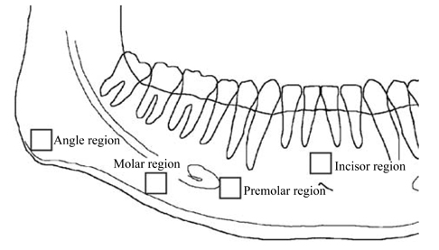
Fig. 1 Locations of region of interest (ROI) selected in the angle, the molar, the premolar and incisor regions.
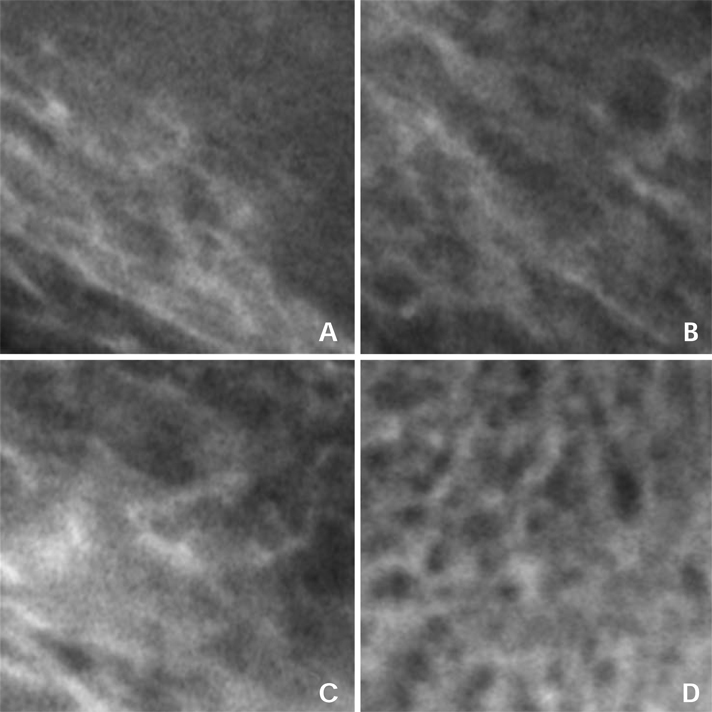
Fig. 2 The images of ROI show the trabecular bone structure. A. Mandibular angle region, B. Mandibular molar region, C. Mandibular premolar region, D. Mandibular incisor region.
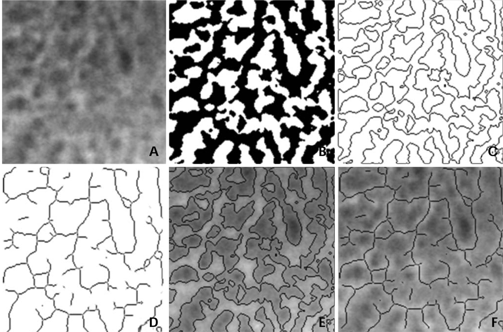
Fig. 3 Image processing procedures. A. Raw image before processing, B. Binary image, C. Outline image, D. Skeletonized image after preprocessing, E. Composition image of raw and outline images, F. Composition image of raw and skeletonized images.
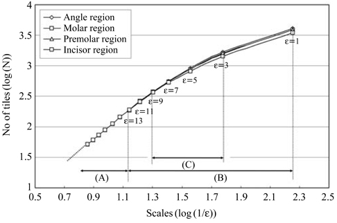
Fig. 4 Mean number of tiles containing any part of the outline against the edge length of the tile or scale from 1 to 49 pixels in 4 anatomical regions. A. A range of scales with local slope of two, B. A range of scales considered for analysis, C. A determined range of scales significant for the fractal dimension.
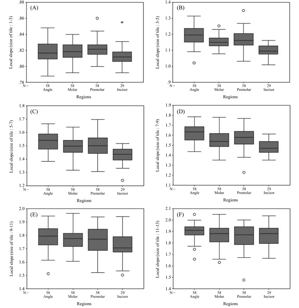
Fig. 5 Box plots show the distribution of local slopes in ranges of scales. A. 1 to 3 pixels, B. 3 to 5 pixels, C. 5 to 7 pixels, D. 7 to 9 pixels, E. 9 to 11 pixels, F. 11 to 13 pixels. No significant differences among the regions in slopes of 1-3, 9-11, and 11-13. There are significant differences among the regions in slopes of 3-5, 5-7, and 7-9 (p<0.05).
Cited by 1 articles
-
Comparison of cone-beam computed tomography and digital panoramic radiography for detecting peri-implant alveolar bone changes using trabecular micro-structure analysis
Guldane Magat, Elif Oncu, Sevgi Ozcan, Kaan Orhan
J Korean Assoc Oral Maxillofac Surg. 2022;48(1):41-49. doi: 10.5125/jkaoms.2022.48.1.41.
Reference
-
1. Mandelbrot BB. The fractal geometry of nature. 1983. New York: WH Freeman.2. Zamir M. On fractal properties of arterial trees. J Theor Biol. 1999. 197:517–526.
Article3. Eblen-Zajjur A, Salas R, Vanegas H. Fractal analysis of spinal dorsal horn neuron discharges by means of sequential fractal dimension D. Comput Biol Med. 1996. 26:87–95.
Article4. Zbilut JP, Mayer-Kress G, Sobotka PA, O'Toole M, Thomas JX Jr. Bifurcations and intrinsic chaotic and 1/f dynamics in an isolated perfused rat heart. Biol Cybern. 1989. 61:371–378.
Article5. Ishibashi A, Aihara K, Kotani M. Chaos in brain and neurons and an analysis on the fractal dimensions. Iyodenshi To Seitai Kogaku. 1988. 26:57–61.6. Majumdar S, Weinstein RS, Prasad RR. Application of fractal geometry techniques to the study of trabecular bone. Med Phys. 1993. 20:1611–1619.
Article7. Pothuaud L, Lespessailles E, Harba R, Jennane R, Royant V, Eynard E, et al. Fractal analysis of trabecular bone texture on radiographs: discriminant value in postmenopausal osteoporosis. Osteoporos Int. 1998. 8:618–625.
Article8. Lin JC, Grampp S, Link T, Kothari M, Newitt DC, Felsenberg D, et al. Fractal analysis of proximal femur radiographs: correlation with biomechanical properties and bone mineral density. Osteoporos Int. 1999. 9:516–524.
Article9. Jiang C, Giger ML, Chinander MR, Martell JM, Kwak S, Favus MJ. Characterization of bone quality using computer-extracted radiographic features. Med Phys. 1999. 26:872–879.
Article10. Majumdar S, Lin J, Link T, Millard J, Augat P, Ouyang X, et al. Fractal analysis of radiographs: assessment of trabecular bone structure and prediction of elastic modulus and strength. Med Phys. 1999. 26:1330–1340.
Article11. Benhamou CL, Poupon S, Lespessailles E, Loiseau S, Jennane R, Siroux V, et al. Fractal analysis of radiographic trabecular bone texture and bone mineral density: two complementary parameters related to osteoporotic fractures. J Bone Miner Res. 2001. 16:697–704.
Article12. Southard TE, Southard KA, Lee A. Alveolar process fractal dimension and postcranial bone density. Oral Surg Oral Med Oral Pathol Oral Radiol Endod. 2001. 91:486–491.
Article13. Ruttimann UE, Webber RL, Hazelrig JB. Fractal dimension from radiographs of peridental alveolar bone. A possible diagnostic indicator of osteoporosis. Oral Surg Oral Med Oral Pathol. 1992. 74:98–110.14. Southard TE, Southard KA, Jakobsen JR, Hillis SL, Najim CA. Fractal dimension in radiographic analysis of alveolar process bone. Oral Surg Oral Med Oral Pathol Oral Radiol Endod. 1996. 82:569–576.
Article15. Hildebolt CF. Osteoporosis and oral bone loss. Dentomaxillofac Radiol. 1997. 26:3–15.
Article16. Lee KI, Choi SC, Park TW, You DS. Fractal dimension calculated from two types of region of interest. Dentomaxillofac Radiol. 1999. 28:284–289.
Article17. Wilding RJ, Slabbert JC, Kathree H, Owen CP, Crombie K, Delport P. The use of fractal analysis to reveal remodelling in human alveolar bone following the placement of dental implants. Arch Oral Biol. 1995. 40:61–72.
Article18. Shrout MK, Roberson B, Potter BJ, Mailhot JM, Hildebolt CF. A comparison of 2 patient populations using fractal analysis. J Periodontol. 1998. 69:9–13.
Article19. Cha SY, Han WJ, Kim EK. Usefulness of fractal analysis for the diagnosis of periodontitis. Korean J Oral Maxillofac Radiol. 2001. 31:35–42.20. Heo MS, Park KS, Lee SS, Choi SC, Koak JY, Heo SJ, et al. Fractal analysis of mandibular bony healing after orthognathic surgery. Oral Surg Oral Med Oral Pathol Oral Radiol Endod. 2002. 94:763–767.
Article21. Law AN, Bollen AM, Chen SK. Detecting osteoporosis using dental radiographs: a comparison of four methods. J Am Dent Assoc. 1996. 127:1734–1742.
Article22. Paumgartner D, Losa G, Weibel ER. Resolution effect on the stereological estimation of surface and volume and its interpretation in terms of fractal dimensions. J Microsc. 1981. 121:51–63.
Article23. Caligiuri P, Giger ML, Favus M. Multifractal radiographic analysis of osteoporosis. Med Phys. 1994. 21:503–508.
Article24. Parkinson IH, Fazzalari NL. Methodological principles for fractal analysis of trabecular bone. J Microsc. 2000. 198:134–142.
Article25. Eriksen EF, Mosekilde L, Melsen F. Trabecular bone resorption depth decreases with age: differences between normal male and females. Bone. 1985. 6:141–146.26. Weinstein RS, Hutson MS. Decreased trabecular width and increased trabecular spacing contribute to bone loss with aging. Bone. 1987. 8:137–142.
Article27. Mosekilde L. Age-related changes in vertebral trabecular bone architecture-assessed by a new method. Bone. 1988. 9:247–250.
Article28. Palle S, Chappard D, Vico L, Riffat G, Alexandre C. Evaluation of the osteoclastic population in iliac crest biopsies from 36 normal subjects: a histoenzymologic and histomorphometric study. J Bone Miner Res. 1989. 4:501–506.
Article29. Moore RJ, Durbridge TC, McNeil PJ, Parkinson IH, Need AG, Vernon-Roberts B. Trabecular spacing in post-menopausal Australian women with and without vertebral fractures. Aust N Z J Med. 1992. 22:269–273.30. Geraets WG, van der Stelt PF. Fractal properties of bone. Dentomaxillofac Radiol. 2000. 29:144–153.
Article31. Geraets WG, Van der Stelt PF, Netelenbos CJ, Elders PJ. A new method for automatic recognition of the radiographic trabecular pattern. J Bone Miner Res. 1990. 5:227–233.
Article32. White SC, Rudolph DJ. Alterations of the trabecular pattern of the jaws in patients with osteoporosis. Oral Surg Oral Med Oral Pathol Oral Radiol Endod. 1999. 88:628–635.
Article
- Full Text Links
-
- Actions
-
Cited
- CITED
-
- Close
- Share
-
- Similar articles
-
- The influence of X ray beam angulation on the fractal analysis of trabecular architecture in human dry mandible using standardized tile counting method
- Effect of exposure time and image resolution on fractal dimension
- Changes in the fractal dimension of peri-implant trabecular bone after loading: a retrospective study
- Change of the fractal dimension according to the decalcification degree and the exposure time in the bovine rib
- Radiologic assessment of bone healing by fractal analysis after the treatment of jaw bone cyst by decompression
