Increase in concentration of soluble HLA-G in high-quality embryos after intracytoplasmic sperm injection
- Affiliations
-
- 1Department of Anatomy, Shahid Beheshti Medical University, Velenjak, Tehran, Iran. hdr@sbmu.ac.ir
- 2Institute of Bioscience, UPM University, Serdang, Selangor, Malaysia.
- 3IVF Center, Taleghani Hospital, Iran.
- 4Cellular and Molecular Research, Shahid Beheshti Medical University, Velenjak, Iran.
- 5Medical University of Tehran, Kargar, Tehran, Iran.
- 6Reproductive Laboratory, Tohoko University, Sendai, Japan.
- KMID: 1447447
- DOI: http://doi.org/10.5115/acb.2011.44.4.331
Abstract
- Non-invasive methods are normally preferred to conventional invasive methods when selecting suitable embryos to improve pregnancy rates after assisted reproduction techniques. One of the most recognized non-invasive methods is to examine the supernatants of embryo culture media. Soluble human leukocyte antigen, class I, G (sHLA-G) antigen is a non-classical class I molecule that has been widely considered as a marker of pregnancy failure or implantation success. In the current study of some Iranian patients, we examined the concentration of sHLA-G at different time points after intracytoplasmic sperm injection and compared the rates to the morphology and quality of the selected embryos. We showed that the concentration of sHLA-G increases over time in high-quality embryos. We conclude that there is a positive relationship between morphology, quality, and sHLA-G concentration. We suggest that this relationship can be used to increase the chance of a successful pregnancy.
MeSH Terms
Figure
-
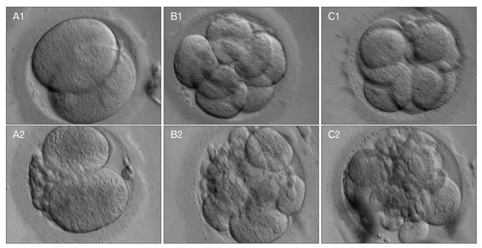
Fig. 1 The morphology of embryos at different time points post-intracytoplasmic sperm injection (ICSI) (×400). (A1) An embryo 48 h post-ICSI. (A2). An embryo 48 h post-ICSI, although the quality was lower compared to A1. (B1) An embryo 72 h post-ICSI. (B2) An embryo 72 h post-ICSI, although the quality was lower compared to B1. (C1) An embryo 72 h post-ICSI at morula stage. (C2) An embryo 72 h post-ICSI at morula stage, although the quality was lower compared to C1.
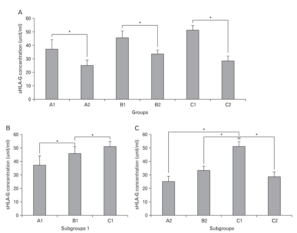
Fig. 2 The concentration of sHLA-G in each group measured by enzyme-linked immunosorbent assay. (A) The concentration in all groups/subgroups. (B) Comparison of the concentration between each subgroup 1. Note that the concentration increased in each group compared to the previous group. (C) Comparison of the concentration between each subgroup 2. Addition of C1 in this figure was to show there was no statistically significant change between subgroups 2. Each data point represents three independent measurements and the error bars indicate the standard error of the mean. sHLA-G, soluble human leukocyte antigen, class I, G. *P<0.05 were referred to as statistically significant differences.
Reference
-
1. Noci I, Fuzzi B, Rizzo R, Melchiorri L, Criscuoli L, Dabizzi S, Biagiotti R, Pellegrini S, Menicucci A, Baricordi OR. Embryonic soluble HLA-G as a marker of developmental potential in embryos. Hum Reprod. 2005. 20:138–146.2. Lessey BA. Embryo quality and endometrial receptivity: lessons learned from the ART experience. J Assist Reprod Genet. 1998. 15:173–176.3. Fisch JD, Rodriguez H, Ross R, Overby G, Sher G. The Graduated Embryo Score (GES) predicts blastocyst formation and pregnancy rate from cleavage-stage embryos. Hum Reprod. 2001. 16:1970–1975.4. Suzumori N, Sugiura-Ogasawara M. Genetic factors as a cause of miscarriage. Curr Med Chem. 2010. 17:3431–3437.5. Rebmann V, Switala M, Eue I, Grosse-Wilde H. Soluble HLA-G is an independent factor for the prediction of pregnancy outcome after ART: a German multi-centre study. Hum Reprod. 2010. 25:1691–1698.6. Ellis SA, Palmer MS, McMichael AJ. Human trophoblast and the choriocarcinoma cell line BeWo express a truncated HLA Class I molecule. J Immunol. 1990. 144:731–735.7. Paul P, Cabestre FA, Ibrahim EC, Lefebvre S, Khalil-Daher I, Vazeux G, Quiles RM, Bermond F, Dausset J, Carosella ED. Identification of HLA-G7 as a new splice variant of the HLA-G mRNA and expression of soluble HLA-G5, -G6, and -G7 transcripts in human transfected cells. Hum Immunol. 2000. 61:1138–1149.8. Rebmann V, Switala M, Eue I, Schwahn E, Merzenich M, Grosse-Wilde H. Rapid evaluation of soluble HLA-G levels in supernatants of in vitro fertilized embryos. Hum Immunol. 2007. 68:251–258.9. Sher G, Keskintepe L, Nouriani M, Roussev R, Batzofin J. Expression of sHLA-G in supernatants of individually cultured 46-h embryos: a potentially valuable indicator of 'embryo competency' and IVF outcome. Reprod Biomed Online. 2004. 9:74–78.10. Hunt JS, Petroff MG, McIntire RH, Ober C. HLA-G and immune tolerance in pregnancy. FASEB J. 2005. 19:681–693.11. Hviid TV, Hylenius S, Lindhard A, Christiansen OB. Association between human leukocyte antigen-G genotype and success of in vitro fertilization and pregnancy outcome. Tissue Antigens. 2004. 64:66–69.12. Moreau P, Paul P, Rouas-Freiss N, Kirszenbaum M, Dausset J, Carosella ED. Molecular and immunologic aspects of the nonclassical HLA class I antigen HLA-G: evidence for an important role in the maternal tolerance of the fetal allograft. Am J Reprod Immunol. 1998. 40:136–144.13. Ng ST, Chang TH, Wu TC. Prediction of the rates of fertilization, cleavage, and pregnancy success by cumulus-coronal morphology in an in vitro fertilization program. Fertil Steril. 1999. 72:412–417.14. Choudhury SR, Knapp LA. Human reproductive failure II: immunogenetic and interacting factors. Hum Reprod Update. 2001. 7:135–160.15. Menicucci A, Noci I, Fuzzi B, Criscuoli L, Scarselli G, Baricordi O, Mattiuz PL. Non-classic sHLA class I in human oocyte culture medium. Hum Immunol. 1999. 60:1054–1057.16. Bamberger AM, Jenatschke S, Schulte HM, Löning T, Bamberger MC. Leukemia inhibitory factor (LIF) stimulates the human HLA-G promoter in JEG3 choriocarcinoma cells. J Clin Endocrinol Metab. 2000. 85:3932–3936.17. Margreiter M, Weghofer A, Kogosowski A, Mahmoud KZ, Feichtinger W. A prospective randomized multicenter study to evaluate the best day for embryo transfer: does the outcome justify prolonged embryo culture? J Assist Reprod Genet. 2003. 20:91–94.18. Riteau B, Rouas-Freiss N, Menier C, Paul P, Dausset J, Carosella ED. HLA-G2, -G3, and -G4 isoforms expressed as nonmature cell surface glycoproteins inhibit NK and antigen-specific CTL cytolysis. J Immunol. 2001. 166:5018–5026.19. Kotze DJ, Hansen P, Keskintepe L, Snowden E, Sher G, Kruger T. Embryo selection criteria based on morphology VERSUS the expression of a biochemical marker (sHLA-G) and a graduated embryo score: prediction of pregnancy outcome. J Assist Reprod Genet. 2010. 27:309–316.20. Alinejad Z, Jafari Shakib R, Forghan-Parast K, Zahiri Z, Sadri H, Nagafi F, Roushan Z. In vitro fertilized embryos do not secrete detectable HLA-G on day two. Iran J Immunol. 2009. 6:195–201.
- Full Text Links
-
- Actions
-
Cited
- CITED
-
- Close
- Share
-
- Similar articles
-
- A Case of Pregnancy from Embryos following ICSI with Frozen-Thawed Testicular Sperms
- Twin Pregnancy and Delivery After Intracytoplasmic Sperm Injection Followed by Calcium Ionophore with Spermatozoa from a Globozoospermic Man: A Case Report
- A Case of Pregnancy from Cryopreserved Embryos following ICSI with Frozen-Thawed Epididymal Sperms
- High mRNA expression of GABA receptors in human sperm with oligoasthenoteratozoospermia and teratozoospermia and its association with sperm parameters and intracytoplasmic sperm injection outcomes
- Physiological intracytoplasmic sperm injection does not improve the quality of embryos: A cross-sectional investigation on sibling oocytes
