KIT-negative Gastrointestinal Stromal Tumor in a Child: A Case Report
- Affiliations
-
- 1Department of Radiology, Wonkwang University School of Medicine and Hospital, Korea. yjyh@wonkwang.ac.kr
- 2Department of Pediatirics, Wonkwang University School of Medicine and Hospital, Korea.
- 3Department of Pathology, Wonkwang University School of Medicine and Hospital, Korea.
- KMID: 1443589
- DOI: http://doi.org/10.3348/jksr.2011.64.1.91
Abstract
- We report here on the imaging findings of the case of KIT-negative gastrointestinal stromal tumor (GIST) in the stomach of a 12-year-old girl. Radiologic studies revealed the presence of a huge exophytic growing mass that originated from the gastric wall and this mass consisted of solid and cystic components on USG, CT and MR. The cystic regions were mainly located at the periphery of the mass and they were revealed to be myxoid degeneration and hemorrhage on histopathologic examination. The tumor consisted of epithelioid and typical spindle cells and they showed negative immunoreactivity for KIT. Although KIT-negative GISTs are rare, they can be considered in the differential diagnosis when a large heterogeneous extraluminal mass that contains solid portions and various degrees of peripheral cystic regions is observed.
Figure
-
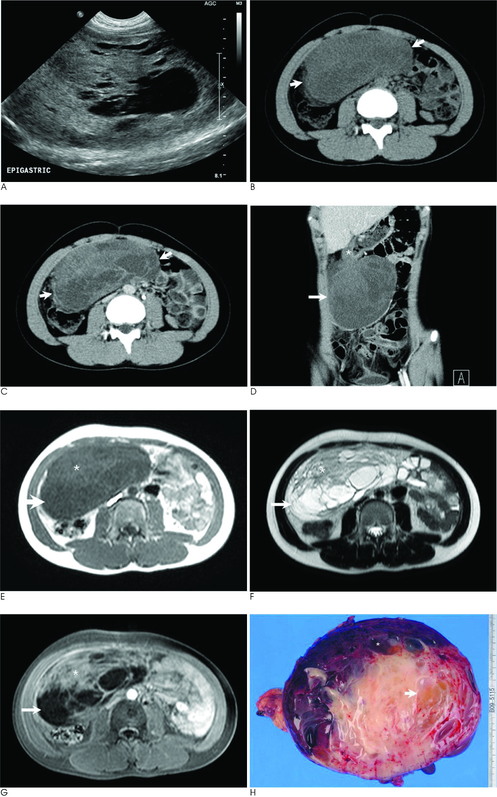
Fig. 1 A 12-year-old girl presented with a KIT-negative gastrointestinal stromal tumor of the stomach. A. Abdominal ultrasonography shows a huge mass with heterogenous echogenicity and the mass is composed of cystic and solid regions. B, C. Axial pre- and post- contrast CT images show a huge mass (arrows) occupying the abdominal cavity. The mass shows enhancing solid and non-enhancing cystic components. D. Coronal contrast-enhanced CT image shows a heterogeneously enhanced exophytic mass (arrow) originating from the gastric wall of the lower body (asterisk). E-G. Axial T1-weighted MR image (E), T2-weighted MR image (F) and contrast-enhanced T1-weighted MR image (G) show a complex mass with cystic and solid regions. The solid regions (asterisk) have intermediate signal intensity on both the T1- and T2-weighted images with slight contrast enhancement on the contrast-enhanced T1-weighted image. The cystic regions (arrow) show low signal intensity on the T1-weighted image and high signal intensity on the T2-weighted image without contrast enhancement on the contrast-enhanced T1-weighted image. H. Photograph of gross pathologic specimen revealed that the tumor had cystic regions that showed myxoid degeneration (arrow) and hemorrhage (asterisk).
Reference
-
1. Prakash S, Sarran L, Socci N, DeMatteo RP, Eisenstat J, Greco AM, et al. Gastrointestinal stromal tumors in children and young adults: a clinicopathologic, molecular, and genomic study of 15 cases and review of the literature. J Pediatr Hematol Oncol. 2005; 27:179–187.2. Kaemmer DA, Otto J, Lassay L, Steinau G, Klink C, Junge K, et al. The Gist of literature on pediatric GIST review of clinical presentation. J Pediatr Hematol Oncol. 2009; 31:108–112.3. Debiec-Rychter M, Wasag B, Stul M, De Wever I, Van Oosterom A, Hagemeijer A, et al. Gastrointestinal stromal tumours (GISTs) negative for KIT (CD117 antigen) immunoreactivity. J Pathol. 2004; 202:430–438.4. Tateishi U, Miyake M, Maeda T, Arai Y, Seki K, Hasegawa T. CT and MRI findings in KIT-weak or KIT-negative atypical gastrointestinal stromal tumors. Eur Radiol. 2006; 16:1537–1543.5. Medeiros F, Corless CL, Duensing A, Hornick JL, Oliveira AM, Heinrich MC, et al. KIT-negative gastrointestinal stromal tumors: proof of concept and therapeutic implications. Am J Surg Pathol. 2004; 28:889–894.6. Miettinen M, Lasota J. Gastrointestinal stromal tumors: review of morphology, molecular pathology, prognosis, and differential diagnosis. Arch Pathol Lab Med. 2006; 130:1466–1478.7. Sakurai S, Hasegawa T, Sakuma Y, Takazawa Y, Motegi A, Nakajima T, et al. Myxoid epithelioid gastrointestinal stromal tumor (GIST) with mast cell infiltrations: a subtype of GIST with mutations of platelet-derived growth factor receptor alpha gene. Hum Pathol. 2004; 35:1223–1230.8. Sandrasegaran K, Rajesh A, Rushing DA, Rydberg J, Akisik FM, Henley JD. Gastrointenstinal stromal tumors: CT and MRI findings. Eur Radiol. 2005; 15:1407–1414.9. Kim HC, Lee JM, Kim SH, Park SH, Lee JW, Lee M, et al. Small gastrointestinal stromal tumours with focal areas of low attenuation on CT: pathological correlation. Clin Radiol. 2005; 60:384–388.10. Suh JS, Abenoza P, Galloway HR, Everson LI, Griffiths HJ. Peripheral (extracranial) nerve tumors: correlation of MR imaging and histologic findings. Radiology. 1992; 183:341–346.
- Full Text Links
-
- Actions
-
Cited
- CITED
-
- Close
- Share
-
- Similar articles
-
- C-Kit-Negative Gastrointestinal Stromal Tumor in the Stomach
- Gastrointestinal stromal tumor with KIT mutation in neurofibromatosis type 1
- Gastrointestinal Stromal Tumor of the Rectum in a Pediatric
- A Case of an Extragastrointestinal Stromal Tumor of the Pelvic Cavity Presenting with Hematuria
- Primary Retroperitoneal Malignant Gastrointestinal Stromal Tumor Mimicking Adrenal Mass
