High-resolution Computed Tomography Findings of H1N1 Influenza-Associated Pneumonia in Korea
- Affiliations
-
- 1Department of Radiology, Chonnam National University Hospital, Chonnam National University, Medical School, Korea. sunaura@hanmail.net
- 2Department of Radiology, Chonnam National University Hwasun Hospital, Chonnam National University, Medical School, Korea.
- 3Department of Internal Medicine, Chonnam National University Hospital, Chonnam National University, Medical School, Korea.
- KMID: 1443538
- DOI: http://doi.org/10.3348/jksr.2011.64.4.351
Abstract
- PURPOSE
To evaluate and compare the high-resolution computed tomography (HRCT) findings of patients with H1N1 influenza-associated pneumonia compared usual community acquired pneumonia (CAP), to determine whether there were any useful common HRCT findings predicting their prognosis.
MATERIALS AND METHODS
HRCT findings of 31 patients (M:F=16:15, mean age 42 yrs) with Influenza A (H1N1) infection were retrospectively reviewed for abnormal HRCT findings and compared to HRCT findings of CAP in matched patients. Patients were matched according to age and sex, from 2009 to January 2010.
RESULTS
The predominant HRCT findings of pneumonia consisted of areas of consolidation and/or ground-glass opacity (GGO) which showed no statistically significant differences when comparing the two groups. However, the abnormalities of H1N1-related pneumonia showed higher bilaterality and multilobar or multisegmental involvement compared with CAP (p<0.05). Internal low attenuation or air-densities in pulmonary infiltration /or lymphadenopathy was observed only in patients with CAP (p<0.05). HRCT findings in 8 patients with poor clinical outcome had bilaterality (p=0.015), multilobar, and multisegmental involvement.
CONCLUSION
The predominant HRCT findings of H1N1-related pneumonia were areas of consolidation and/or GGO. In addition, H1N1-related pneumonia showed higher bilaterality or multilobar/multisegmental involvement compared with CAP. The patients who presented bilaterality had a worse clinical outcome.
MeSH Terms
Figure
-

Fig. 1 H1N1 influenza-associated pneumonia in a 31-year-old woman with fever. A chest HRCT scan shows subsegmental consolidation (black arrow) in right middle lobe and diffuse bronchial wall thickening (white arrow) in both lower lobes, and small centrilobular nodules with linear branching opacities (arrow heads) in right lower lobe.
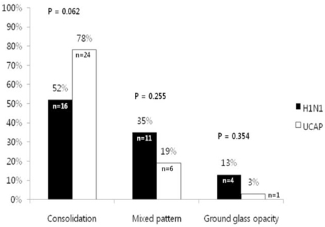
Fig. 2 HRCT patterns of abnormal parenchymal opacities in H1N1 associated pneumonia (black bar) and usual community acquired pneumonia (white bar) are consolidation, mixed pattern, or GGO. Each pattern of parenchymal opacities does not show statistically significant difference.

Fig. 3 H1N1 influenza-associated pneumonia in a 51-year-old woman with cough. A chest HRCT scan shows multifocal consolidations (black arrow) with mild surrounding ground glass attenuation (arrowhead) in both lower lobes with posterior predominance.
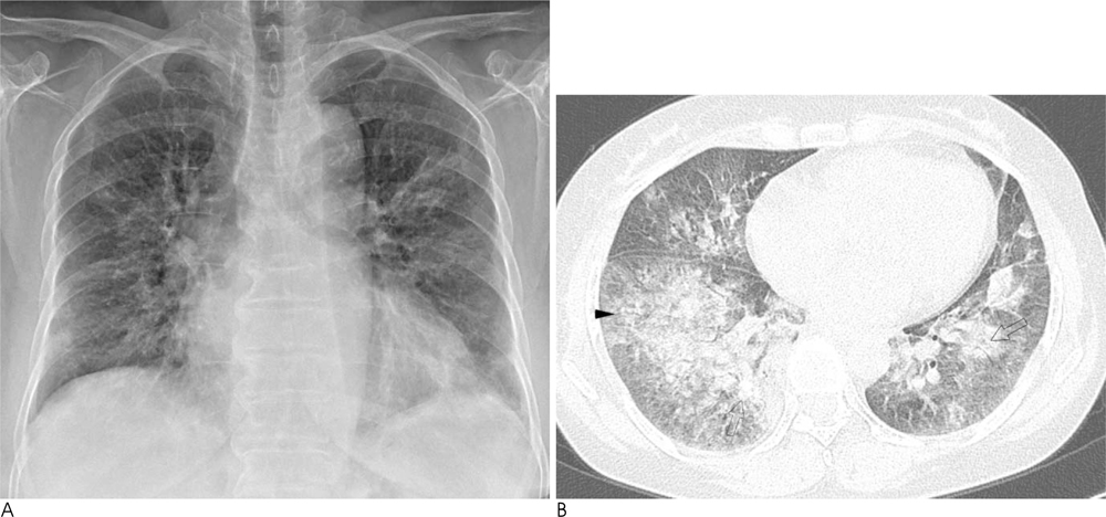
Fig. 4 H1N1 influenza-associated pneumonia in a 50-year-old woman with fever. An initial chest radiograph (A) shows multiple patchy areas of increased opacities in both lungs. Chest HRCT scan (B) shows multiple patchy areas of consolidations (open arrows in B) and ground glass attenuations (arrowhead in B) in both lungs.
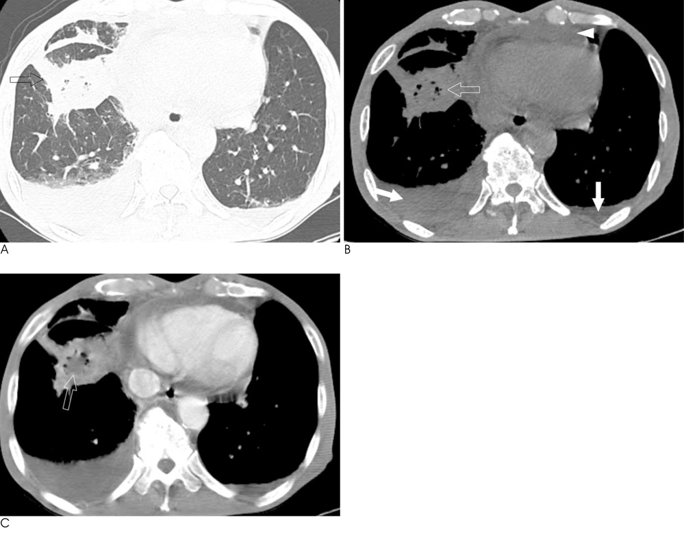
Fig. 5 Usual community acquired pneumonia in 79-year-old male with fever and general weakness. Chest CT scans (A-C) show consolidation in right lower lobe (open arrow in A) containing areas of mottled air densities (open arrow in B) and necrotic low attenuation (open arrow in C, postcontrast enhanced mediastinal window setting). Bilateral pleural (white arrows in B) and pericardial effusion (white arrowhead in B) are also noted.
Reference
-
1. Perez-Padilla R, de la Rosa-Zamboni D, Ponce de Leon S, Hernandez M, Quinones-Falconi F, Bautista E, et al. Pneumonia and respiratory failure from swine-origin influenza A (H1N1) in mexico. N Engl J Med. 2009; 361:680–689.2. Centers for disease control and prevention. Novel H1N1 flu: Background on the situation. [Internet]. Atlanta: The Association;updated 2009 May 25. Available from: http://www.Cdc.Gov/h1n1flu/background.Html.3. WHO. Global alert and response: Pandemic (H1N1) 2009 : Update 100. [Internet]. Geneva: World health organization;cited 2011 Jan 31. Availabe from: http://www.who.int/csr/don/2010_05_14/en/.4. WHO. Media centre: H1N1 in post-pandemic period [Internet]. Geneva: World health organization;cited 2011 Jan 31. Available from: Http://www.who.Int/mediacentre/news/statements/2010/h1n1_vpc_20100810/en/.5. Kim EA, Lee KS, Primack SL, Yoon HK, Byun HS, Kim TS, et al. Viral pneumonias in adults: radiologic and pathologic findings. Radiographics. 2002; 22:S137–S149.6. Agarwal PP, Cinti S, Kazerooni EA. Chest radiographic and CT findings in novel swine-origin influenza A (H1N1) virus (S-OIV) infection. AJR Am J Roentgenol. 2009; 193:1488–1493.7. Ajlan AM, Quiney B, Nicolaou S, Muller NL. Swine-origin influenza A (H1N1) viral infection: radiographic and CT findings. AJR Am J Roentgenol. 2009; 193:1494–1499.8. Lee EY, McAdam AJ, Chaudry G, Fishman MP, Zurakowski D, Boiselle PM. Swine-origin influenza A (H1N1) viral infection in children: initial chest radiographic findings. Radiology. 2010; 254:934–941.9. Mollura DJ, Asnis DS, Crupi RS, Conetta R, Feigin DS, Bray M, et al. Imaging findings in a fatal case of pandemic swine-origin influenza A (H1N1). AJR Am J Roentgenol. 2009; 193:1500–1503.10. Tanaka N, Matsumoto T, Kuramitsu T, Nakaki H, Ito K, Uchisako H, et al. High resolution CT findings in community-acquired pneumonia. J Comput Assist Tomogr. 1996; 20:600–608.11. WHO. CDC protocol of realtime rtpcr for influenza A (H1N1) 2009. [Internet]. Genova: World health organization;update 2009 Apr 30. Available from: http://www.who.int/csr/resources/publications/swineflu/realtimeptpcr/en/.12. Ramsdell J, Narsavage GL, Fink JB. Management of communityacquired pneumonia in the home: an american college of chest physicians clinical position statement. Chest. 2005; 127:1752–1763.13. Hansell DM, Bankier AA, MacMahon H, McLoud TC, Muller NL, Remy J. Fleischner society: glossary of terms for thoracic imaging. Radiology. 2008; 246:697–722.14. Elicker BM, Schwartz BS, Liu C, Chen EC, Miller SA, Chiu CY, et al. Thoracic CT findings of novel influenza A (H1N1) infection in immunocompromised patients. Emerg Radiol. 2010; 17:299–307.15. Li P, Su DJ, Zhang JF, Xia XD, Sui H, Zhao DH. Pneumonia in novel swine-origin influenza A (H1N1) virus infection: high-resolution CT findings. Eur J Radiol. 2010; Forthcoming.16. Soto-Abraham MV, Soriano-Rosas J, Diaz-Quinonez A, Silva-Pereyra J, Vazquez-Hernandez P, Torres-Lopez O, et al. Pathological changes associated with the 2009 H1N1 virus. N Engl J Med. 2009; 361:2001–2003.17. Jain S, Kamimoto L, Bramley AM, Schmitz AM, Benoit SR, Louie J, et al. Hospitalized patients with 2009 H1N1 influenza in the united states, april-june 2009. N Engl J Med. 2009; 361:1935–1944.18. Pittet LA, Hall-Stoodley L, Rutkowski MR, Harmsen AG. Influenza virus infection decreases tracheal mucociliary velocity and clearance of streptococcus pneumoniae. Am J Respir Cell Mol Biol. 2010; 42:450–460.19. Dominguez-Cherit G, Lapinsky SE, Macias AE, Pinto R, Espinosa-Perez L, de la Torre A, et al. Critically ill patients with 2009 influenza A (H1N1) in mexico. JAMA. 2009; 302:1880–1887.20. Aviram G, Bar-Shai A, Sosna J, Rogowski O, Rosen G, Weinstein I, et al. H1N1 influenza: initial chest radiographic findings in helping predict patient outcome. Radiology. 2010; 255:252–259.21. Guo HH, Sweeney RT, Regula D, Leung AN. Best cases from the afip: fatal 2009 influenza A (H1N1) infection, complicated by acute respiratory distress syndrome and pulmonary interstitial emphysema. Radiographics. 2010; 30:327–333.
- Full Text Links
-
- Actions
-
Cited
- CITED
-
- Close
- Share
-
- Similar articles
-
- Follow-up Aspects of Influenza A (H1N1) Virus-Associated Pneumonia: the Role of High-Resolution Computed Tomography in the Evaluation of the Recovery Phase
- Radiologic Findings of Influenza A (H1N1) Pneumonia: Report of Two Cases
- Influenza Associated Pneumonia
- A Case of Novel Influenza A (H1N1) Virus Pneumonia Complicated Pnemomediastinum and Subcutenous Emphysema
- RE: Pediatric Novel Influenza A (H1N1) Virus Infection: the Imaging Findings
