Analysis of Complex Coronary Plaque in Multidetector Computed Tomography: Comparison with Conventional Coronary Angiography
- Affiliations
-
- 1Department of Radiology, Soonchunhyang University Hospital Bucheon, Korea. dhk0827@schmc.ac.kr
- 2Department of Internal Medicine, Soonchunhyang University Hospital, Korea.
- 3Department of Anesthesiology and Pain Medicine, Asan Medical Center, Korea.
- 4Department of Internal Medicine, Soonchunhyang University Hospital Bucheon, Korea.
- KMID: 1443532
- DOI: http://doi.org/10.3348/jksr.2011.64.4.309
Abstract
- PURPOSE
To delineate complex plaque morphology in patients with stable angina using coronary computed tomographic angiography (CTA).
MATERIALS AND METHODS
36 patients with complex plaques proven by conventional coronary angiography (CAG), who had taken CTA for evaluation of typical angina, were enrolled in this study. Intravascular ultrasonography (IVUS) was performed in 14 patients (16 lesions). We compared CTA with CAG for plaque features and analyzed vascular cutoff, intraluminal filling defect in a patent vessel, irregularity of plaque, and ulceration. Also, the density of plaque was evaluated on CTA.
RESULTS
CAG and CTA showed complex morphology in 44 cases (100%) and 34 cases, (77%), respectively, with features including abrupt vessel cutoff (27 vs. 16%, kappa= 0.57), intraluminal filling defect (32 vs. 30%, kappa= 0.77), irregularity (75 vs. 52%, kappa= 0.52), and ulceration (16 vs. 11%, kappa= 0.60). CTA indicated that the complex lesions were hypodense (mean = 66 +/- 21 Houndsfield Units).
CONCLUSION
CTA is a very accurate and useful non-invasive imaging modality for evaluating complex plaque in patients with typical angina.
MeSH Terms
Figure
-
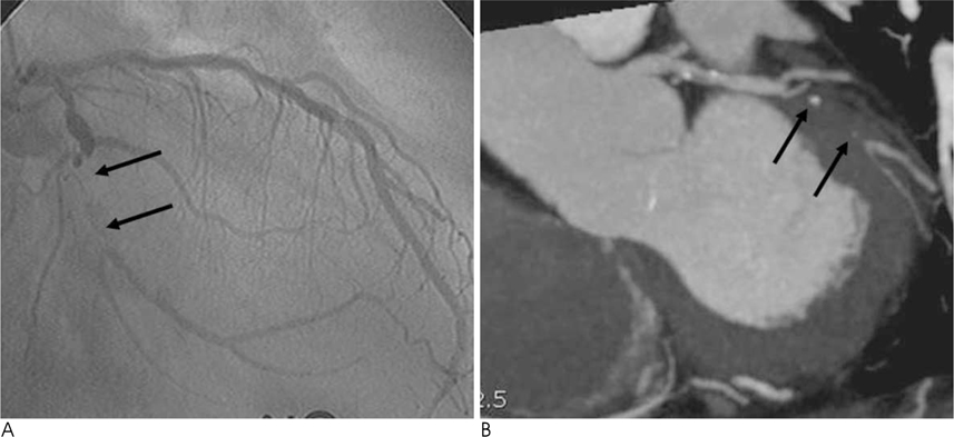
Fig. 1 A 71-year-old man with chest pain. Invasive coronary angiography (A) shows LCX plaque characterized by bulky, lucent lesion with occlusion of distal LCX (arrows). CTA image (B) reveals similar morphology characterized by a bulky, hypodense plaque (arrows) with contrast filling distally in distal segment of LCX.

Fig. 2 A 63-year-old man with unstable ruptured RCA lesion. Invasive coronary angiography (A) documents filling defect, complex plaque severely narrowing the proximal segment of RCA (arrows). CTA image (B) shows a concordant bulky, eccentric and hypodense filling defect in proximal segment of RCA (arrow).

Fig. 3 A 78-year-old man with acute chest pain. Coronary angiogram (A) demonstrates severely irregular, stenotic lesion (black arrow) and occlusion (white arrow) in the proximal and middle segment of RCA, respectively. The CTA image (B) shows eccentric and hypodense plaque with irregular margin (arrow) compared to invasive coronary angiography.

Fig. 4 A 69-year-old man with atypical chest pain over 7 days duration. Invasive coronary angiography (A) shows hazy irregular lesion (dark arrow), with punctuate zone of ulceration (arrow) in middle segment of LAD. CTA (B) demonstrates the LAD lesion as irregular and eccentric low density plaque with intra-plaque contrast penetration indicative of ulceration (arrow).
Reference
-
1. Lee SG, Lee CW, Hong MK, Kim JJ, Park SW, Park SJ. Change of multiple complex coronary plaques in patients with acute myocardial infarction: a study with coronary angiography. Am Heart J. 2004; 147:281–286.2. Hong YJ, Mintz GS, Kim SW, Okabe T, Bui AB, Pichard AD, et al. Impact of plaque rupture and elevated C-reactive protein on clinical outcome in patients with acute myocardial infarction: an intravascular ultrasound study. J Invasive Cardiol. 2008; 20:428–435.3. Hong MK, Mintz GS, Lee CW, Kin YH, Lee SW, Song JM, et al. Comparison of coronary plaque rupture between stable angina and acute myocardial infarction: a three-vessel intravascular ultrasound study in 235 patients. Circulation. 2004; 110:928–933.4. Hoffmann MH, Shi H, Schmitz BL, Schmid FT, Lieberknecht M, Schulze R, et al. Noninvasive coronary angiography with multislice computed tomography. JAMA. 2005; 293:2471–2478.5. Herzog C, Zwerner PL, Doll JR, Nielsen CD, Nguyen SA, Savino G, et al. Significant coronary artery stenosis: comparison on perpatient and per-vessel or per-segment basis at 64-section CT angiography. Radiology. 2007; 244:112–120.6. Meijboom WB, Weustink AC, Pugliese F, van Mieghem CA, Mollet NR, van Pelt N, et al. Comparison of diagnostic accuracy of 64-slice computed tomography coronary angiography in women versus men with angina pectoris. Am J Cardiol. 2007; 100:1532–1537.7. Goldstein JA, Gallagher MJ, O'Neill WW, Ross MA, O'Neil BJ, Raff GL. A randomized controlled trial of multi-Slice coronary computed tomography for evaluation of acute chest pain. J Am Coll Cardiol. 2007; 49:863–871.8. Hoffmann U, Nagurnev JT, Moselewski F, Pena A, Ferencik M, Chae CU, et al. Coronary multidetector computed tomography in the assessment of patients with acute chest pain. Circulation. 2006; 114:2251–2260.9. Noda M, Takagi A, Kuwatsuru R, Mitsuhashi N, Kasanuki H. Prognostic significance of multiple-detector computed tomography in conjunction with TIMI risk score for patients with non-ST elevation acute coronary syndrome. Heart Vessels. 2008; 23:161–166.10. Rehr R, Disciascio G, Vetrover G, Cowley M. Angiographic morphology of coronary artery stenosis in prolonged rest angina: evidence of intracoronary thrombosis. J Am Coll Cardiol. 1989; 14:1429–1437.11. Qiao JH, Fishbein MC. The severity of coronary atherosclerosis at sites of plaque rupture with occlusive thrombosis. J Am Coll Cardiol. 1991; 17:1138–1142.12. Mintz GS, Nissen SE, Anderson WD, Bailey SR, Erbel R, Fitzgerald PJ, et al. American College of Cardiology clinical expert consensus document on standards for acquisition, measurement and reporting of intravascular ultrasound studies (IVUS): a report of the American College of Cardiology task force on clinical expert consensus documents. J Am Coll Cardiol. 2001; 37:1478–1492.13. Kotani J, Mintz GS, Castagna MT, Pinnow E, Berzingi CO, Bui AB, et al. Intravascular ultrasound analysis of infarct-related and non-infarct-related arteries in patients who presented with an acute myocardial infarction. Circulation. 2003; 107:2889–2893.14. Raff GL, Goldstein JA. Coronary angiography by computed tomography: coronary imaging evolves. J Am Coll Cardiol. 2007; 49:1830–1833.15. Sun J, Zhang Z, Lu B, Yu W, Yang Y, Zhou Y, et al. Identification and quantification of coronary atherosclerotic plaques: a comparison of 64-MDCT and intravascular ultrasound. AJR Am J Roentgenol. 2008; 190:748–754.16. Dragu R, Kerner A, Gruberg L, Rispler S, Lessick J, Ghersin E, et al. Angiographically uncertain left main coronary artery narrowings: correlation with multidetector computed tomography and intravascular ultrasound. Int J Cardiovasc Imaging. 2008; 24:557–563.17. Pohle K, Achenbach S, Macneill B, Ropers D, Ferencik M, Moselewski F, et al. Characterization of non-calcified coronary atherosclerotic plaque by multi-detector row CT: comparison to IVUS. Atherosclerosis. 2007; 190:174–180.18. Komatsu S, Hirayama A, Omori Y, Ueda Y, Mizote I, Fujisawa Y, et al. Detection of coronary plaque by computed tomography with a novel plaque analysis system, 'Plaque Map', and comparison with intravascular ultrasound and angioscopy. Circ J. 2005; 69:72–77.19. Achenbach S, Moselewski F, Ropers D, Ferencik M, Hoffmann U, MacNeill B, et al. Detection of calcified and noncalcified coronary atherosclerotic plaque by contrast-enhanced, submillimeter multidetector spiral computed tomography: a segment-based comparison with intravascular ultrasound. Circulation. 2004; 109:14–17.20. Kunimasa T, Sato Y, Sugi K, Moroi M. Evaluation of multislice computed tomography of atherosclerotic coronary artery plaques in non-culprit, remote coronary arteries of patients with acute coronary syndrome. Circ J. 2005; 69:1346–1351.21. Caussin C, Ohanessian A, Ghostine S, Jacq L, Lancelin B, Dambrin G, et al. Characterization of vulnerable nonstenotic plaque with 16-slice computed tomography compared with intravascular ultrasound. Am J Cardiol. 2004; 94:99–104.22. Goldstein JA, Demetriou D, Grines CL, Pica M, Shoukfeh M, O'Neill WW. Multiple complex coronary plaques in patients with acute myocardial infarction. N Engl J Med. 2000; 343:915–922.23. Little WC, Applegate RJ. The shadows leave a doubt-the angiographic recognition of vulnerable coronary artery plaques. J Am Coll Cardiol. 1999; 33:1362–1364.24. Schroeder S, Kopp AF, Baumbach A, Meisner C, Kuettner A, Georg C, et al. Noninvasive detection and evaluation of atherosclerotic coronary plaques with multislice computed tomography. J Am Coll Cardiol. 2001; 37:1430–1435.25. Leber AW, Knez A, Becker A, Becker C, von Ziegler F, Nikolaou K, et al. Accuracy of multidetector spiral computed tomography in identifying and differentiating the composition of coronary atherosclerotic plaques: a comparative study with intracoronary ultrasound. J Am Coll Cardiol. 2004; 43:1241–1247.26. Klein LW, Calvin JE. Unstable angina: pathogenetic mechanisms, coronary angiographic observations, risk stratification, and therapeutic implications. Heart Dis. 1999; 1:19–28.
- Full Text Links
-
- Actions
-
Cited
- CITED
-
- Close
- Share
-
- Similar articles
-
- Coronary CT Angiography
- A Single Coronary Artery: Right Coronary Artery Originating From the Distal Left Circumflex Artery
- Giant coronary aneurysm caused by Kawasaki disease: consistency between catheter angiography and electrocardiogram gated dual-source computed tomography angiography
- Anomalous Origin of the Left Circumflex Coronary Artery from the Right Sinus of Valsalva Identified by Imaging with Multidetector Computed Tomography
- Napkin-Ring Sign on Coronary Computed Tomography Angiography-Tiered Enhancement of Coronary Lumen and Plaque
