Aberrant Cervical Thymus Mimicking Thyroid on Ultrasonography: A Case Report
- Affiliations
-
- 1Department of Radiology, Jeju National University Hospital, Jeju National University School of Medicine, Jeju, Korea. jkcontrast@naver.com
- 2Department of Surgery, Jeju National University Hospital, Jeju National University School of Medicine, Jeju, Korea.
- KMID: 1439531
- DOI: http://doi.org/10.3348/jksr.2012.67.4.231
Abstract
- Aberrant cervical thymus is rarely reported in adults. We report a case of solid aberrant cervical thymus in a 27-year-old female, which was found incidentally on ultrasonography for the evaluation of the thyroid cancer. On ultrasonography, the lesion was found between the left thyroid and common carotid artery without any remarkable interface echo, and had similar echogenicity to the thyroid. The lesion extended to the upper pole of the left thyroid.
Figure
-
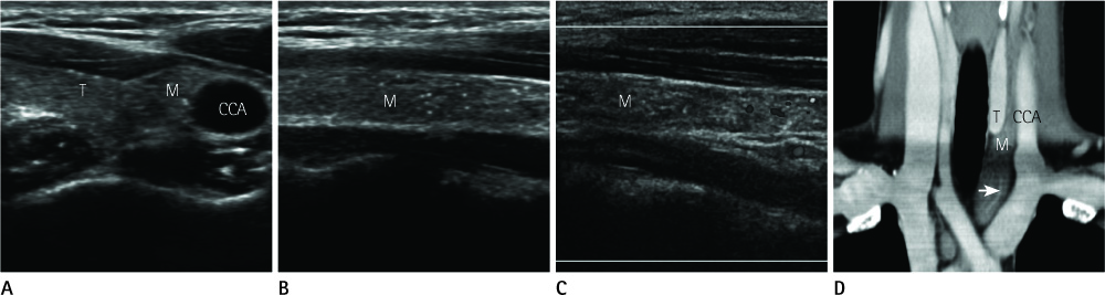
Fig. 1 Ultrasonography reveals a solid mass between the left thyroid gland and CCA, which has elongated tubular shape and similar echogenicity to the thyroid parenchyma (A: transverse image, B: longitudinal image). There is no remarkable interface echo to the thyroid parenchyma. The lesion has multiple internal echogenic dots (A, B). Doppler ultrasonography reveals internal blood flow (C). Post-contrast coronal reconstructed CT scan shows about 7 cm, elongated tubular shaped mass, located between the left thyroid and CCA, and extends from the upper pole level of the left thyroid to the superior mediastinum (D). Several enhancing vascular structures (arrow) are noted in the mass (D). Note.-CCA = common carotid artery, M = mass, T = thyroid
Reference
-
1. Curé JK, Tagge EP, Richardson MS, Mulvihill DM. MR of cystic aberrant cervical thymus. AJNR Am J Neuroradiol. 1995. 16:1124–1127.2. Nowak PA, Zarbo RJ, Jacobs JR. Aberrant solid cervical thymus. Ear Nose Throat J. 1988. 67:670673676–677.3. Sturm-O'Brien AK, Salazar JD, Byrd RH, Popek EJ, Giannoni CM, Friedman EM, et al. Cervical thymic anomalies--the Texas Children's Hospital experience. Laryngoscope. 2009. 119:1988–1993.4. Song I, Yoo SY, Kim JH, Hong E, Yoon HK. Aberrant cervical thymus: imaging and clinical findings in 13 children. Clin Radiol. 2011. 66:38–42.5. Conwell LS, Batch JA. Aberrant cervical thymus mimicking a cervical mass. J Paediatr Child Health. 2004. 40:579–580.6. Costa NS, Laor T, Donnelly LF. Superior cervical extension of the thymus: a normal finding that should not be mistaken for a mass. Radiology. 2010. 256:238–242.7. Han BK, Yoon HK, Suh YL. Thymic ultrasound. II. Diagnosis of aberrant cervical thymus. Pediatr Radiol. 2001. 31:480–487.8. Koumanidou C, Vakaki M, Theophanopoulou M, Koutrouvelis H, Savvidou D, Pitsoulakis G, et al. Aberrant thymus in infants: sonographic evaluation. Pediatr Radiol. 1998. 28:987–989.9. Fitoz S, Atasoy C, Türköz E, Gümüş D, Erden I, Akyar S. Sonographic findings in ectopic cervical thymus in an infant. J Clin Ultrasound. 2001. 29:523–526.10. Lewis CT. Ectopic thymus of the neck--report of three examples in children. Postgrad Med J. 1975. 51:38–40.
- Full Text Links
-
- Actions
-
Cited
- CITED
-
- Close
- Share
-
- Similar articles
-
- Thymoma Arising from Aberrant Cervical Thymus
- Intrathyroidal thymic tissue mimicking a malignant thyroid nodule in a 4-year-old child
- Intraabdominal Heterotopic Thymus: Report of an autopsy case
- A Case of Aberrant Cervical Thymus in a One-year-old Boy
- Spindle Epithelial Tumor with Thymus-Like Differentiation of the Thyroid Gland: A Case Report with Ultrasonography and CT Features, Cytological Findings and Histopathological Results
