Spindle Epithelial Tumor with Thymus-Like Differentiation of the Thyroid Gland: A Case Report with Ultrasonography and CT Features, Cytological Findings and Histopathological Results
- Affiliations
-
- 1Department of Radiology, Busan Paik Hospital, Inje University College of Medicine, Busan, Korea. netisss@hanmail.net
- 2Department of Pathology, Busan Paik Hospital, Inje University College of Medicine, Busan, Korea.
- KMID: 2355992
- DOI: http://doi.org/10.3348/jksr.2016.75.5.384
Abstract
- Spindle epithelial tumor with thymus-like differentiation (SETTLE) of the thyroid gland is a very rare tumor. It is believed to originate from ectopic thymus tissue within the thyroid gland or from branchial pouch remnants that differentiate along the thymic line. A few reports of SETTLE have been presented, but to the best of our knowledge, there is no case report in which detailed preoperative imaging features of SETTLE have been described. In addition, there are no case reports of SETTLE in Korean patients. Thus, we report a case of SETTLE with detailed preoperative ultrasonography and computed tomography features, cytological findings and histopathological results.
Figure
-
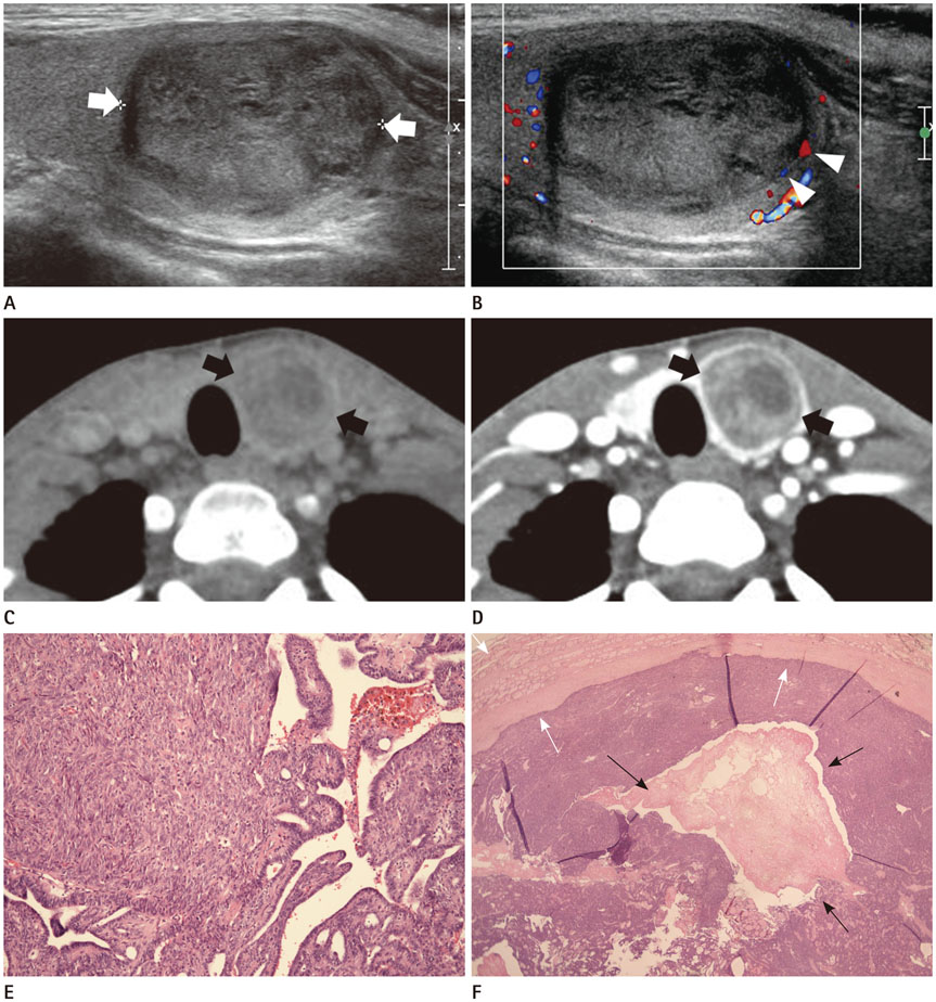
Fig. 1 A 19-year-old woman with spindle epithelial tumor with thymus-like differentiation of the thyroid gland. A, B. Longitudinal gray-scale (A) and longitudinal color Doppler (B) sonograms show a heterogeneous solid thyroid nodule (arrows) in the left lobe with benign findings including smooth margin with a hypoechoic halo, oval shape, isoechogenicity, and peripheral vascularity (arrowheads). C, D. On non-enhanced axial (C) and contrast-enhanced axial (D) neck CT images, the left thyroid nodule (arrows) shows heterogeneous low attenuation, no calcification, smooth margin, and heterogeneous enhancement. A less enhancing central portion of the left thyroid nodule is also noted. There are no other malignant findings such as adjacent soft tissue invasion or suspicious lymphadenopathy. E. A histopathological section of the left thyroid nodule shows a biphasic neoplasm with predominant spindle cells and a minor glandular mucous component (hematoxylin and eosin stain; original magnification, × 200). F. The mass is encapsulated by a thick fibrous capsule (white arrows). There is a pseudocyst (black arrows) composed of a large cystic space and fibrinous material deposition in the central portion of the left thyroid nodule (hematoxylin and eosin stain; original magnification, × 10).
Reference
-
1. Chan JK, Rosai J. Tumors of the neck showing thymic or related branchial pouch differentiation: a unifying concept. Hum Pathol. 1991; 22:349–367.2. Magnata Filho LA, Bordallo MA, Pessoa CH, Corbo R, Bulzico DA, Dias FL, et al. Thyroid spindle epithelial tumor with thymus-like differentiation (SETTLE): case report and review. Arq Bras Endocrinol Metabol. 2010; 54:657–662.3. Erickson ML, Tapia B, Moreno ER, McKee MA, Kowalski DP, Reyes-Múgica M. Early metastasizing spindle epithelial tumor with thymus-like differentiation (SETTLE) of the thyroid. Pediatr Dev Pathol. 2005; 8:599–606.4. Cheuk W, Jacobson AA, Chan JK. Spindle epithelial tumor with thymus-like differentiation (SETTLE): a distinctive malignant thyroid neoplasm with significant metastatic potential. Mod Pathol. 2000; 13:1150–1155.5. Kloboves-Prevodnik V, Jazbec J, Us-Krasovec M, Lamovec J. Thyroid spindle epithelial tumor with thymus-like differentiation (SETTLE): is cytopathological diagnosis possible? Diagn Cytopathol. 2002; 26:314–319.6. Mote DG, Satyanarayana V. Spindle epithelial tumor with thymus-like element of the thyroid gland. World J Endoc Surg. 2012; 4:74–76.7. Kim DW, Park JS, In HS, Choo HJ, Ryu JH, Jung SJ. Ultrasound-based diagnostic classification for solid and partially cystic thyroid nodules. AJNR Am J Neuroradiol. 2012; 33:1144–1149.8. Llamas-Gutierrez FJ, Falcon-Escobedo R, De Anda-Gonzalez J, Angeles-Angeles A. Spindle epithelial tumor with thymus-like differentiation of the thyroid (SETTLE): report of two cases (one associated with a parathyroid adenoma). Ann Diagn Pathol. 2013; 17:217–221.9. Kim DW, Jung SJ, Baek HJ. Computed tomography features of benign and malignant solid thyroid nodules. Acta Radiol. 2015; 56:1196–1202.10. Lee S, Han BK, Ko EY, Oh YL, Choe JH, Shin JH. The ultrasonography features of hyalinizing trabecular tumor of the thyroid are more consistent with its benign behavior than cytology or frozen section readings. Thyroid. 2011; 21:253–259.
- Full Text Links
-
- Actions
-
Cited
- CITED
-
- Close
- Share
-
- Similar articles
-
- Spindle epithelial tumor with thymus-like differentiation of the thyroid in a 70-year-old man
- Carcinoma Showing Thymus-Like Differentiation (CASTLE) of the Thyroid Gland: A case report
- Cytologic Findings of Thyroid Carcinoma Showing Thymus-like Differentiation: A Case Report
- Cytological Features of a Lymphoepithelial Cyst Collected from Fine Needle Aspiration of the Thyroid Gland That Mimicked Papillary Thyroid Carcinoma: A Case Report
- Aberrant Cervical Thymus Mimicking Thyroid on Ultrasonography: A Case Report
