J Korean Fract Soc.
2012 Jan;25(1):58-63. 10.12671/jkfs.2012.25.1.58.
Radiation Exposure Over the Course of a Year from an Image Intensifier in the Orthopaedic Operating Room
- Affiliations
-
- 1Department of Orthopaedic Surgery, Gospel Hospital, Kosin University College of Medicine, Busan, Korea. jyujin2001@kosin.ac.kr
- KMID: 1434112
- DOI: http://doi.org/10.12671/jkfs.2012.25.1.58
Abstract
- PURPOSE
To measure the annual radiation exposure of staff in the orthopaedic surgical room.
MATERIALS AND METHODS
From January 2010 to December 2010, we measured the radiation exposure of a tumor surgeon, spine surgeon, trauma surgeon, six residents, and six scrub nurses. Radiation was monitored with the use of thermoluminescent dosimeters placed on the chest under the lead apron. The annual dose of radiation exposure was compared to the maximum yearly permissible dose (20 mSv). During the study period, the trauma surgeon made a deliberate effort to minimize the radiation time and maintain a distance of 1 m from the image intensifier.
RESULTS
The annual exposure levels were 0.04 mSv (radiation time, 34 min 50 s), 0.08 mSv (151 min 46 s), and 0.12 mSv (135 min 27 s) for the tumor surgeon, trauma surgeon, and spine surgeon, respectively. The mean exposure was 0.0146 mSv (range, 0.4~0.39 mSv) for the residents and 0.06 mSv (range, 0.04~0.13 mSv) for the scrub nurses. Overall, the annual radiation exposure was 0.2~1.95% of the maximal yearly permissible dose. Despite the longer period of radiation exposure, the trauma surgeon was exposed to a lower dose of radiation than the spine surgeon.
CONCLUSION
The annual radiation exposure of a trauma surgeon can be reduced by a deliberate effort to decrease exposure time and maintain a distance of at least 1 m from the image intensifier.
Keyword
MeSH Terms
Figure
-
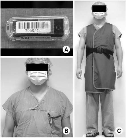
Fig. 1 (A~C) The thermoluminescent dorsimeter (TLD) which is placed on the chest under the lead apron, is used for the monitoring of radiation.
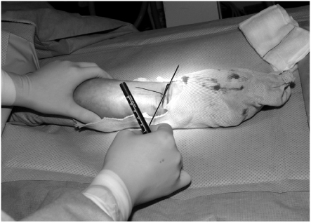
Fig. 2 The drawing on the skin and provisional K-wire fixations are done to minimize the missed image.
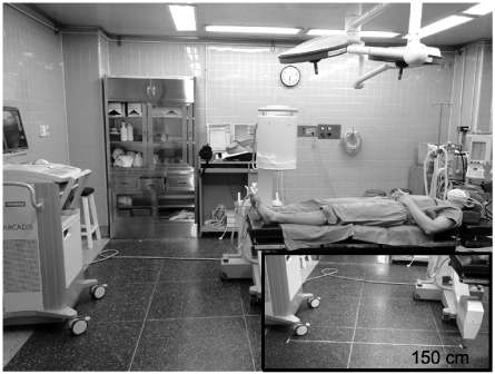
Fig. 3 The monitor of image intensifier is pushed against wall in foot's direction.

Fig. 4 In order to maintaine a safe distance (100 cm) from the image intensifier, the orthopaedic surgeon takes a step away from operating table before (A) and after (B).
Reference
-
1. ARCADIS VARIC, User's manual for technical specifications. 2010. Germany: Siemens Co.
Article2. Bar-On E, Weigl DM, Becker T, Katz K, Konen O. Intraoperative C-arm radiation affecting factors and reduction by an intervention program. J Pediatr Orthop. 2010. 30:320–323.
Article3. Fuchs M, Schmid A, Eiteljörge T, Modler M, Stürmer KM. Exposure of the surgeon to radiation during surgery. Int Orthop. 1998. 22:153–156.
Article4. Giachino AA, Cheng M. Irradiation of the surgeon during pinning of femoral fractures. J Bone Joint Surg Br. 1980. 62:227–229.5. Giannoudis PV, McGuigan J, Shaw DL. Ionising radiation during internal fixation of extracapsular neck of femur fractures. Injury. 1998. 29:469–472.6. International Commission on Radiological Protection. 1990 recommendations of the international commission on radiological protection. 1991. 21:Ann ICRP;1–201.
Article7. Kim JW, Kim JJ. Radiation exposure to the orthopaedic surgeon during fracture surgery. J Korean Orthop Assoc. 2010. 45:107–113.
Article8. Levin PE, Schoen RW Jr, Browner BD. Radiation exposure to the surgeon during closed interlocking intramedullary nailing. J Bone Joint Surg Am. 1987. 69:761–766.
Article9. Mahaisavariya B, Songcharoen P, Riansuwan K. Radiation scattering to the primary surgeon during closed locked femoral nailing. J Med Assoc Thai. 2005. 88:252–255.
Article10. Maxon HR, Thomas SR, Saenger EL, Buncher CR, Kereiakes JG. Ionizing irradiation and the induction of clinically significant disease in the human thyroid gland. Am J Med. 1977. 63:967–978.
Article11. Mehlman CT, DiPasquale TG. Radiation exposure to the orthopaedic surgical team during fluoroscopy: "how far away is far enough?". J Orthop Trauma. 1997. 11:392–398.
Article12. Müller LP, Suffner J, Wenda K, Mohr W, Rommens PM. Radiation exposure to the hands and the thyroid of the surgeon during intramedullary nailing. Injury. 1998. 29:461–468.
Article13. Noordeen MH, Shergill N, Twyman RS, Cobb JP, Briggs T. Hazard of ionizing radiation to trauma surgeons: reducing the risk. Injury. 1993. 24:562–564.
Article
- Full Text Links
-
- Actions
-
Cited
- CITED
-
- Close
- Share
-
- Similar articles
-
- Radiation Exposure from Fluoroscopy during Orthopaedic Surgical Procedures
- Radiation exposure in diagnostic radiology
- Pattern and degree of radiation exposure during endovascular surgery performed using a mobile C-arm or in a hybrid room
- Percutaneous Ablation of Osteoid Osteoma under Image Intensifier Guidance: A Case Report
- A New Shielding Curtain for Protection of Intraoperative Radiation During Minimally Invasive Spine Surgery
