Intracranial Pial Arteriovenous Fistula Presenting with Hemorrhage: A Case Report
- Affiliations
-
- 1Department of Neurosurgery, Seoul National University Bundang Hospital, Department of Neurosurgery, Seoul National University College of Medicine, Seoul, Korea. wanoh@snu.ac.kr
- KMID: 1431285
- DOI: http://doi.org/10.7461/jcen.2012.14.4.305
Abstract
- Intracranial pial arteriovenous fistula (AVF) is a rare cerebrovascular malformation, which has a single or multiple arterial connections to a single venous channel without intervening nidus, and is different from arteriovenous malformation (AVM). We report on a case of a surgically treated pial AVF. A 15-year-old girl with an altered mental state was brought to our hospital. Computed tomography (CT) showed a subcortical hematoma of approximately 24 ml in her right temporal lobe. Cerebral angiography showed an AVF supplied by the right middle cerebral artery with early drainage into the Sylvian vein and the vein of Labbe. She underwent surgical treatment with feeding artery obliteration using a clip and hematoma removal. The patient was discharged without neurologic deficits. Despite the rarity of pial AVF, for correct diagnosis and treatment, neurosurgeons should recognize this condition. Pial AVF can be managed simply by disconnection of the shunt by surgery or endovascular treatment, and a good result can be achieved.
Keyword
MeSH Terms
Figure
-

Fig. 1 Initial Computed Tomography (CT) and Magnetic Resonance Imaging (MRI) of the patient. [A] CT angiography shows a suspicious abnormal vessel (arrow) around the hematoma on the right temporal lobe. [B] An Arterio-Venous Malformation (AVM) nidus is not observed using enhanced MRI.
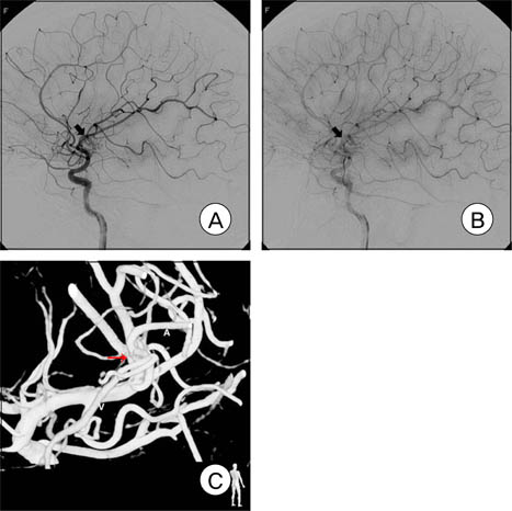
Fig. 2 Preoperative cerebral angiography of the patient. [A] A possible Arterio-Venous Fistula (AVF) (arrow) is observed around the right Middle Cerebral Artery (MCA) bifurcation. [B] The AVF drains into the Sylvian vein and the vein of Labbe in the early venous phase. [C] A 3 Dimensional (D) image shows the direct connection (arrow) of the right MCA (A) and the surrounding vein (V).
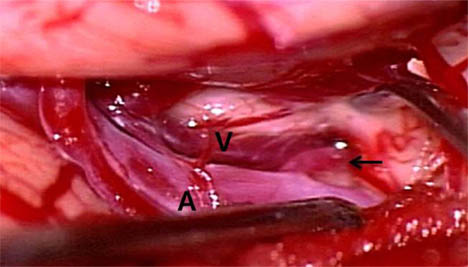
Fig. 3 Intraoperative photographs. Black arrow indicates the AVF consisting of the feeding artery (A) and drainage vein (V) show in preoperative angiography.

Fig. 4 Cerebral angiography one week postoperatively. [A] The early draining venous flow through the Sylvian vein and the vein of Labbe disappeared. [B] A 3D image shows that the direct connection of the artery and vein is disconnected (arrow).
Cited by 1 articles
-
Pial Arteriovenous Fistula with Giant Varices: Report of Two Cases with Good Surgical Outcome
Morteza Faghih Jouibari, Mehdi Zeinali Zadeh, Masoud Khadivi, Alireza Khoshnevisan, Keisan Moazzeni, Sina Abdollahzade
J Cerebrovasc Endovasc Neurosurg. 2014;16(2):98-103. doi: 10.7461/jcen.2014.16.2.98.
Reference
-
1. Yang WH, Lu MS, Cheng YK, Wang TC. Pial arteriovenous fistula: a review of literature. Br J Neurosurg. 2011. 10. 25(5):580–585.
Article2. Halbach VV, Higashida RT, Hieshima GB, Hardin CW, Dowd CF, Barnwell SL. Transarterial occlusion of solitary intracerebral arteriovenous fistulas. AJNR Am J Neuroradiol. 1989. Jul-Aug. 10(4):747–752.3. Yamashita K, Ohe N, Yoshimura SI, Iwama T. Intracranial pial arteriovenous fistula. Neurol Med Chir (Tokyo). 2007. 47(12):550–554.4. Lasjaunias P, Manelfe C, Chiu M. Angiographic architecture of intracranial vascular malformations and fistulas-pretherapeutic aspects. Neurosurg Rev. 1986. 9(4):253–263.5. Giller CA, Batjer HH, Purdy P, Walker B, Mathews D. Interdisciplinary evaluation of cerebral hemodynamics in the treatment of arteriovenous fistulae associated with giant varices. Neurosurgery. 1994. 10. 35(4):778–782. discussion 782-4.
Article6. Halbach VV, Higashida RT, Hieshima GB, Norman D. Normal perfusion pressure breakthrough occurring during treatment of carotid and vertebral fistulas. AJNR Am J Neuroradiol. 1987. Sep-Oct. 8(5):751–756.7. Almeida GM, Shibata MK. Hemispheric arteriovenous fistulae with giant venous dilation. Childs Nerv Syst. 1990. 06. 6(4):216–219.
Article8. Hoh BL, Putman CM, Budzik RF, Ogilvy CS. Surgical and endovascular flow disconnection of intracranial pial single-channel arteriovenous fistulae. Neurosurgery. 2001. 11. 49(6):1351–1363. discussion 1363-4.
Article9. Talamonti G, Versari PP, D'Aliberti G, Villa F, Fontana RA, Collice M. Complex arteriovenous fistula of the brain in an infant. Case report. J Neurosurg Sci. 1997. 12. 41(4):337–341.10. Garcia-Monaco R, De Victor D, Mann C, Hannedouche A, Terbrugge K, Lasjaunias P. Congestive cardiac manifestations from cerebrocranial arteriovenous shunts. Endovascular management in 30 children. Childs Nerv Syst. 1991. 02. 7(1):48–52.11. Drake CG. Cerebral arteriovenous malformations: considerations for and experience with surgical treatment in 166 cases. Clin Neurosurg. 1979. 26:145–208.
Article12. Wang YC, Wong HF, Yeh YS. Intracranial pial arteriovenous fistulas with single-vein drainage. Report of three cases and review of the literature. J Neurosurg. 2004. 02. 100:2 Suppl Pediatrics. 201–205.13. Nelson PK, Niimi Y, Lasjaunias P, Berenstein A. Endovascular embolization of congenital intracranial pial arteriovenous fistulas. Neuroimaging Clin N Am. 1992. 2(2):309–317.14. Antunes JL, DiGiacinto GV, Michelsen WJ. Giant hemispheric arteriovenous fistula in an infant. Surg Neurol. 1977. 01. 7(1):45–48.15. Aoki N, Sakai T, Oikawa A. Intracranial arteriovenous fistula manifesting as progressive neurological deterioration in an infant: case report. Neurosurgery. 1991. 04. 28(4):619–622. discussion 622-3.
Article16. Bendok BR, Getch CC, Frederiksen J, Batjer HH. Resection of a large arteriovenous fistula of the brain using low-flow deep hypothermic cardiopulmonary bypass: technical case report. Neurosurgery. 1999. 04. 44(4):888–890. discussion 890-1.
Article17. Black KL, Rubin JM, Chandler WF, McGillicuddy JE. Intraoperative color-flow Doppler imaging of AVM's and aneurysms. J Neurosurg. 1988. 04. 68(4):635–639.
Article18. Gilsbach JM, Hassler WE. Intraoperative Doppler and real time sonography in neurosurgery. Neurosurg Rev. 1984. 7(2-3):199–208.
Article
- Full Text Links
-
- Actions
-
Cited
- CITED
-
- Close
- Share
-
- Similar articles
-
- Successful Treatment of Intracranial Small Pial Single-Channel Arteriovenous Fistula Using N-butyl Cyanoacrylate: Report of 2 Cases
- Intracranial Pial Arteriovenous Fistula Presenting as Brain Hemorrhage in Newborn Infants
- Congenital Intracranial Pial Arteriovenous Fistula Complicated with Congestive Heart Failure in Neonate: A Case Report
- Embolization of Cerebral Pial Arteriovenous Fistula Under Balloon-assisted Flow Control Using NBCA: a Case Report
- Borden Type I Sigmoid Sinus Dural Arteriovenous Fistula Presenting as Subarachnoid Hemorrhage from a Feeding Artery Aneurysm of the Anterior Inferior Cerebellar Artery: A Case Report
