Opportunities for 2-[18F] Fluoro-2-Deoxy-D-Glucose PET/CT in Cervical-Vaginal Neuroendocrine Carcinoma: Case Series and Literature Review
- Affiliations
-
- 1Department of Nuclear Medicine, Taichung Veterans General Hospital, Taichung 404, Taiwan.
- 2Graduate Institute of Clinical Medicine Science and School of Medicine, College of Medicine, China Medical University, Taichung 404, Taiwan. d10040@mail.cmuh.org.tw
- 3Department of Radiation Oncology, China Medical University Hospital, Taichung 404, Taiwan.
- 4Department of Nuclear Medicine and PET Center, China Medical University Hospital, Taichung 404, Taiwan.
- KMID: 1397507
- DOI: http://doi.org/10.3348/kjr.2012.13.6.760
Abstract
OBJECTIVE
Neuroendocrine cervical carcinoma is a rare subtype of cervical cancer. These tumors exhibit an aggressive behavior with early regional lymph node and distant metastases. The purpose of our study was to describe five cases of neuroendocrine cervical-vaginal carcinoma and to discuss the potential of the 2-[18F] fluoro-2-deoxy-D-glucose positron emission tomography/computed tomography (18F-FDG PET/CT) scan for the detection of this rare malignancy.
MATERIALS AND METHODS
Five cases of cervical-vaginal neuroendocrine tumor were retrospectively collected, during a two year (from September 2009 to August 2011) period in our hospital. The clinical staging distributions were International Federation of Gynecology and Obstetrics (FIGO) stage IB2 (1 of 5), stage IIA (3 of 5) and stage IVA (1 of 5).
RESULTS
Two cases (cases 1 and 4) were restaged after 18F-FDG PET/CT scan in the initial staging process. Post-treatment 18F-FDG PET/CT scans, in three patients, revealed positive findings for tumor recurrence or lymph node metastases. Two patients (cases 2 and 3) died of tumor within two years.
CONCLUSION
18F-FDG PET/CT scan is a useful tool in cervical-vaginal neuroendocrine tumor. In its initial staging, the 18F-FDG PET/CT scan may help assess the possible nodal involvement or early hematogeneous spreading. We can also use the 18F-FDG PET/CT to detect local recurrence and to evaluate the treatment response after clinical manipulation.
MeSH Terms
Figure
-
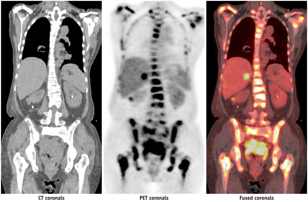
Fig. 1 2-[18F] fluoro-2-deoxy-D-glucose positron emission tomography/computed tomography images showed multiple hypermetabolic areas in skeleton (including axial bones and long bones of four limbs), retroperitoneal lymph nodes, liver, lung, uterine cervix, and posterior wall of urinary bladder. Renal cortex, hepatic parenchyma, and soft tissue uptake were faintly visualized. PET = positron emission tomography
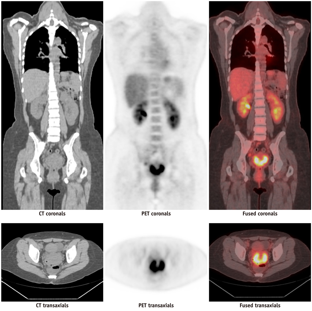
Fig. 2 2-[18F] fluoro-2-deoxy-D-glucose positron emission tomography/computed tomography images showed lesion in uterine cervix and no distal metastases. PET = positron emission tomography
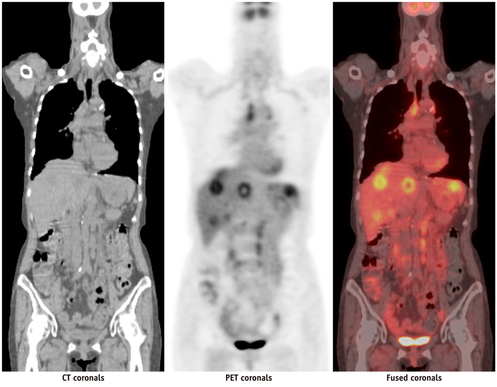
Fig. 3 2-[18F] fluoro-2-deoxy-D-glucose positron emission tomography/computed tomography (18F-FDG PET/CT) images after radical hysterectomy showed multiple 18F-FDG-avid lesions in mediastinum, liver and left kidney, which were consistent with metastases.
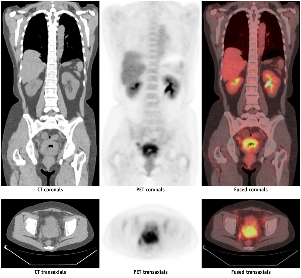
Fig. 4 2-[18F] fluoro-2-deoxy-D-glucose positron emission tomography/computed tomography (18F-FDG PET/CT) images showed huge 18F-FDG-avid mass in posterior pelvis, which was compatible with cervical cancer. Several hypermetabolic lesions in bilateral iliac and retroperitoneal regions were demonstrated, which were mostly related to metastatic lymph nodes. Incidentally, hypermetabolic lesion in left breast was also noted. Biopsy of this lesion showed metastatic neuroendocrine carcinoma.
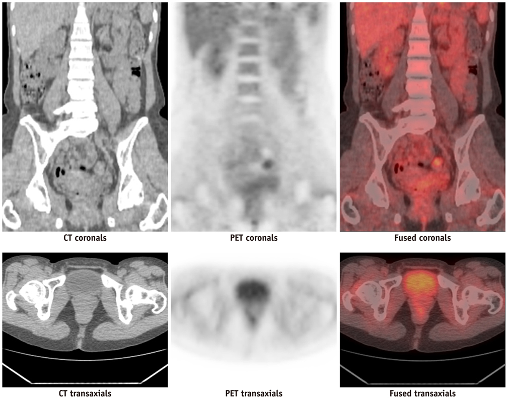
Fig. 5 2-[18F] fluoro-2-deoxy-D-glucose positron emission tomography/computed tomography images showed lesion in vagina. PET = positron emission tomography
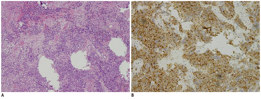
Fig. 6 This is 28-year-old woman. Histopathological examination (× 100) of tumor showed infiltration of blue round cells with vesicular nuclei, scanty cytoplasma, high nuclear/cytoplasma ratio and extensive necrosis arranged in nest on hematoxylin and eosin stain (A); and positive immunohistochemical stain for synaptophysin (B). These features confirmed diagnosis of neuroendocrine carcinoma.
Reference
-
1. Oberg K, Castellano D. Current knowledge on diagnosis and staging of neuroendocrine tumors. Cancer Metastasis Rev. 2011. 30:Suppl 1. 3–7.2. Albores-Saavedra J, Gersell D, Gilks CB, Henson DE, Lindberg G, Santiago H, et al. Terminology of endocrine tumors of the uterine cervix: results of a workshop sponsored by the College of American Pathologists and the National Cancer Institute. Arch Pathol Lab Med. 1997. 121:34–39.3. Sato Y, Shimamoto T, Amada S, Asada Y, Hayashi T. Large cell neuroendocrine carcinoma of the uterine cervix: a clinicopathological study of six cases. Int J Gynecol Pathol. 2003. 22:226–230.4. Krivak TC, McBroom JW, Sundborg MJ, Crothers B, Parker MF. Large cell neuroendocrine cervical carcinoma: a report of two cases and review of the literature. Gynecol Oncol. 2001. 82:187–191.5. Reig Castillejo A, Membrive Conejo I, Foro Arnalot P, Rodríguez de Dios N, Algara López M. Neuroendocrine small cell carcinoma of the uterine cervix. Clin Transl Oncol. 2010. 12:512–513.6. McCusker ME, Coté TR, Clegg LX, Tavassoli FJ. Endocrine tumors of the uterine cervix: incidence, demographics, and survival with comparison to squamous cell carcinoma. Gynecol Oncol. 2003. 88:333–339.7. Wang KL, Yang YC, Wang TY, Chen JR, Chen TC, Chen HS, et al. Neuroendocrine carcinoma of the uterine cervix: a clinicopathologic retrospective study of 31 cases with prognostic implications. J Chemother. 2006. 18:209–216.8. Chung HH, Nam BH, Kim JW, Kang KW, Park NH, Song YS, et al. Preoperative [18F]FDG PET/CT maximum standardized uptake value predicts recurrence of uterine cervical cancer. Eur J Nucl Med Mol Imaging. 2010. 37:1467–1473.9. Nakamura K, Okumura Y, Kodama J, Hongo A, Kanazawa S, Hiramatsu Y. The predictive value of measurement of SUVmax and SCC-antigen in patients with pretreatment of primary squamous cell carcinoma of cervix. Gynecol Oncol. 2010. 119:81–86.10. Yilmaz M, Adli M, Celen Z, Zincirkeser S, Dirier A. FDG PET-CT in cervical cancer: relationship between primary tumor FDG uptake and metastatic potential. Nucl Med Commun. 2010. 31:526–531.11. Jhingran A, Levenback C. Katz VL, Lentz GM, Lobo RA, Gershenson DM, editors. Malignagnt diseases of the cervix: microinvasive and invasive carcinoma: diagnosis and management. Comprehensive Gynecology. 2007. 5th ed. Philadelpia, PA: Mosby Elsevier.12. Henriksen E. The lymphatic spread of carcinoma of the cervix and of the body of the uterus; a study of 420 necropsies. Am J Obstet Gynecol. 1949. 58:924–942.13. Yang DH, Kim JK, Kim KW, Bae SJ, Kim KH, Cho KS. MRI of small cell carcinoma of the uterine cervix with pathologic correlation. AJR Am J Roentgenol. 2004. 182:1255–1258.14. Okamoto Y, Tanaka YO, Nishida M, Tsunoda H, Yoshikawa H, Itai Y. MR imaging of the uterine cervix: imaging-pathologic correlation. Radiographics. 2003. 23:425–445. quiz 534-535.15. Yen TC, Ng KK, Ma SY, Chou HH, Tsai CS, Hsueh S, et al. Value of dual-phase 2-fluoro-2-deoxy-d-glucose positron emission tomography in cervical cancer. J Clin Oncol. 2003. 21:3651–3658.16. Unger JB, Lilien DL, Caldito G, Ivy JJ, Charrier A, Bellaire B. The prognostic value of pretreatment 2-[18F]-fluoro-2-deoxy-D-glucose positron emission tomography scan in women with cervical cancer. Int J Gynecol Cancer. 2007. 17:1062–1067.17. Waggoner SE. Cervical cancer. Lancet. 2003. 361:2217–2225.18. Trinh XB, Bogers JJ, Van Marck EA, Tjalma WA. Treatment policy of neuroendocrine small cell cancer of the cervix. Eur J Gynaecol Oncol. 2004. 25:40–44.19. Belhocine TZ. 18F-FDG PET imaging in posttherapy monitoring of cervical cancers: from diagnosis to prognosis. J Nucl Med. 2004. 45:1602–1604.20. Pallardy A, Bodet-Milin C, Oudoux A, Campion L, Bourbouloux E, Sagan C, et al. Clinical and survival impact of FDG PET in patients with suspicion of recurrent cervical carcinoma. Eur J Nucl Med Mol Imaging. 2010. 37:1270–1278.
- Full Text Links
-
- Actions
-
Cited
- CITED
-
- Close
- Share
-
- Similar articles
-
- Clinical Usefulness of 18F 2-Fluoro-2-Deoxy-D-Glucose Positron Emission Tomography Scan in the Diagnosis of Ampullary Carcinoma
- Recurrent Follicular Dendritic Cell Sarcoma of the Parotid Gland Imaged with 18F-FDG PET/CT
- Tubulocystic Carcinoma of the Kidney with Bone Metastasis: A Case Report
- Kikuchi-Fujimoto disease mimicking malignant lymphoma with 2-[18F]fluoro-2-deoxy-D-glucose PET/CT in children
- Primary Renal Leiomyosarcoma Presenting with Subcutaneous and Osseous Metastases: Staging and Follow-Up with 18F-FDG PET/CT
