Korean J Radiol.
2012 Aug;13(4):417-424. 10.3348/kjr.2012.13.4.417.
Measurement of Intra-Fraction Displacement of the Mediastinal Metastatic Lymph Nodes Using Four-Dimensional CT in Non-Small Cell Lung Cancer
- Affiliations
-
- 1Department of Radiation Oncology, Shandong Cancer Hospital & Institute, Jinan 250117, China. lijianbin@msn.com
- KMID: 1383853
- DOI: http://doi.org/10.3348/kjr.2012.13.4.417
Abstract
OBJECTIVE
To measure the intra-fraction displacements of the mediastinal metastatic lymph nodes by using four-dimensional CT (4D-CT) in non-small cell lung cancer (NSCLC).
MATERIALS AND METHODS
Twenty-four patients with NSCLC, who were to be treated by using three dimensional conformal radiation therapy (3D-CRT), underwent a 4D-CT simulation during free breathing. The mediastinal metastatic lymph nodes were delineated on the CT images of 10 phases of the breath cycle. The lymph nodes were grouped as the upper, middle and lower mediastinal groups depending on the mediastinal regions. The displacements of the center of the lymph node in the left-right (LR), anterior-posterior (AP), and superior-inferior (SI) directions were measured.
RESULTS
The mean displacements of the center of the mediastinal lymph node in the LR, AP, and SI directions were 2.24 mm, 1.87 mm, and 3.28 mm, respectively. There were statistically significant differences between the displacements in the SI and LR, and the SI and AP directions (p < 0.05). For the middle and lower mediastinal lymph nodes, the displacement difference between the AP and SI was statistically significant (p = 0.005; p = 0.015), while there was no significant difference between the LR and AP directions (p < 0.05).
CONCLUSION
The metastatic mediastinal lymph node movements are different in the LR, AP, and SI directions in patients with NSCLC, particularly for the middle and lower mediastinal lymph nodes. The spatial non-uniform margins should be considered for the metastatic mediastinal lymph nodes in involved-field radiotherapy.
Keyword
MeSH Terms
-
Adult
Aged
Aged, 80 and over
Carcinoma, Non-Small-Cell Lung/*radiography/radiotherapy
Contrast Media/diagnostic use
Female
Four-Dimensional Computed Tomography/*methods
Humans
Iohexol/analogs & derivatives/diagnostic use
Lung Neoplasms/*radiography/radiotherapy
Lymphatic Metastasis/*radiography
Male
Mediastinum/radiography
Middle Aged
Radiographic Image Interpretation, Computer-Assisted
Statistics, Nonparametric
Figure
-

Fig. 1 Four-dimensional computed tomography image of 0% phase showing contours of 2R lymph node. These contours are drawn in all of 10 phases and mapped to current image. A. Transverse view. B. Sagittal view. C. Coronal view.
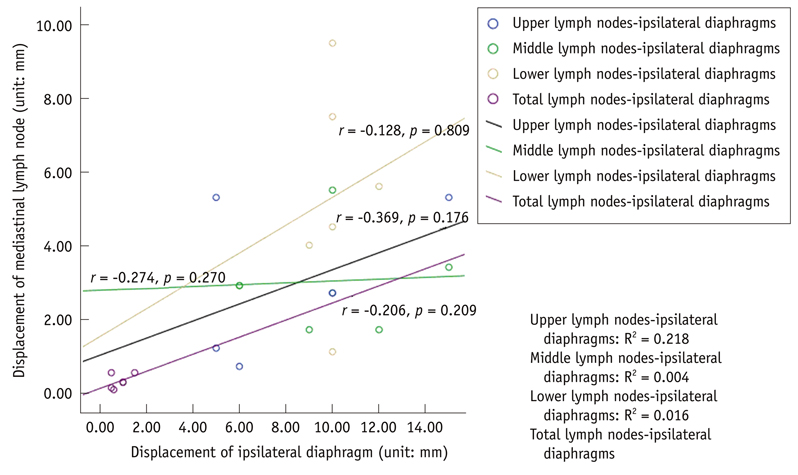
Fig. 2 Correlation between mediastinal lymph node displacement and ipsilateral diaphragm dome movement in superior-inferior direction.
Reference
-
1. Fidler MJ, Kim AW, Zusag T, Bonomi P. Treatment of locally advanced non-small cell lung cancer. Clin Adv Hematol Oncol. 2009. 7:455–464. 479–480.2. Wagner TD, Yang GY. The role of chemotherapy and radiation in the treatment of locally advanced non-small cell lung cancer (NSCLC). Curr Drug Targets. 2010. 11:67–73.3. Emami B, Mirkovic N, Scott C, Byhardt R, Graham MV, James Andras E, et al. The impact of regional nodal radiotherapy (dose/volume) on regional progression and survival in unresectable non-small cell lung cancer: an analysis of RTOG data. Lung Cancer. 2003. 41:207–214.4. Liengswangwong V, Bonner JA. Point: the potential importance of elective nodal irradiation in the treatment of non-small cell lung cancer. Semin Radiat Oncol. 2000. 10:308–314.5. Fernandes AT, Shen J, Finlay J, Mitra N, Evans T, Stevenson J, et al. Elective nodal irradiation (ENI) vs. involved field radiotherapy (IFRT) for locally advanced non-small cell lung cancer (NSCLC): a comparative analysis of toxicities and clinical outcomes. Radiother Oncol. 2010. 95:178–184.6. Rengan R, Rosenzweig KE, Venkatraman E, Koutcher LA, Fox JL, Nayak R, et al. Improved local control with higher doses of radiation in large-volume stage III non-small-cell lung cancer. Int J Radiat Oncol Biol Phys. 2004. 60:741–747.7. Yuan S, Sun X, Li M, Yu J, Ren R, Yu Y, et al. A randomized study of involved-field irradiation versus elective nodal irradiation in combination with concurrent chemotherapy for inoperable stage III nonsmall cell lung cancer. Am J Clin Oncol. 2007. 30:239–244.8. Rosenzweig KE, Sura S, Jackson A, Yorke E. Involved-field radiation therapy for inoperable non small-cell lung cancer. J Clin Oncol. 2007. 25:5557–5561.9. Nakayama H, Satoh H, Kurishima K, Ishikawa H, Tokuuye K. High-dose conformal radiotherapy for patients with stage III non-small-cell lung carcinoma. Int J Radiat Oncol Biol Phys. 2010. 78:645–650.10. Ekberg L, Holmberg O, Wittgren L, Bjelkengren G, Landberg T. What margins should be added to the clinical target volume in radiotherapy treatment planning for lung cancer? Radiother Oncol. 1998. 48:71–77.11. Saunders MI, Dische S, Barrett A, Parmar MK, Harvey A, Gibson D. Randomised multicentre trials of CHART vs conventional radiotherapy in head and neck and non-small-cell lung cancer: an interim report. CHART Steering Committee. Br J Cancer. 1996. 73:1455–1462.12. Shih HA, Jiang SB, Aljarrah KM, Doppke KP, Choi NC. Internal target volume determined with expansion margins beyond composite gross tumor volume in three-dimensional conformal radiotherapy for lung cancer. Int J Radiat Oncol Biol Phys. 2004. 60:613–622.13. Low D. 4D imaging and 4D radiation therapy: a New Era of therapy design and delivery. Front Radiat Ther Oncol. 2011. 43:99–117.14. Jenkins P, Salmon C, Mannion C. Analysis of the movement of calcified lymph nodes during breathing. Int J Radiat Oncol Biol Phys. 2005. 61:329–334.15. van Sörnsen de Koste JR, Lagerwaard FJ, Nijssen-Visser MR, Schuchhard-Schipper R, Joosten H, Senan S. What margins are necessary for incorporating mediastinal nodal mobility into involved-field radiotherapy for lung cancer? Int J Radiat Oncol Biol Phys. 2002. 53:1211–1215.16. Sher DJ, Wolfgang JA, Niemierko A, Choi NC. Quantification of mediastinal and hilar lymph node movement using four-dimensional computed tomography scan: implications for radiation treatment planning. Int J Radiat Oncol Biol Phys. 2007. 69:1402–1408.17. Keall P. 4-dimensional computed tomography imaging and treatment planning. Semin Radiat Oncol. 2004. 14:81–90.18. Low DA, Nystrom M, Kalinin E, Parikh P, Dempsey JF, Bradley JD, et al. A method for the reconstruction of four-dimensional synchronized CT scans acquired during free breathing. Med Phys. 2003. 30:1254–1263.19. Lu W, Parikh PJ, El Naqa IM, Nystrom MM, Hubenschmidt JP, Wahab SH, et al. Quantitation of the reconstruction quality of a four-dimensional computed tomography process for lung cancer patients. Med Phys. 2005. 32:890–901.20. Rietzel E, Pan T, Chen GT. Four-dimensional computed tomography: image formation and clinical protocol. Med Phys. 2005. 32:874–889.21. Keall PJ, Starkschall G, Shukla H, Forster KM, Ortiz V, Stevens CW, et al. Acquiring 4D thoracic CT scans using a multislice helical method. Phys Med Biol. 2004. 49:2053–2067.22. Mountain CF, Dresler CM. Regional lymph node classification for lung cancer staging. Chest. 1997. 111:1718–1723.23. Chapet O, Kong FM, Quint LE, Chang AC, Ten Haken RK, Eisbruch A, et al. CT-based definition of thoracic lymph node stations: an atlas from the University of Michigan. Int J Radiat Oncol Biol Phys. 2005. 63:170–178.24. Pantarotto JR, Piet AH, Vincent A, van Sörnsen de Koste JR, Senan S. Motion analysis of 100 mediastinal lymph nodes: potential pitfalls in treatment planning and adaptive strategies. Int J Radiat Oncol Biol Phys. 2009. 74:1092–1099.25. Liu HH, Balter P, Tutt T, Choi B, Zhang J, Wang C, et al. Assessing respiration-induced tumor motion and internal target volume using four-dimensional computed tomography for radiotherapy of lung cancer. Int J Radiat Oncol Biol Phys. 2007. 68:531–540.26. Li FX, Li JB, Zhang YJ, Liu TH, Tian SY, Xu M, et al. Comparison of the planning target volume based on three-dimensional CT and four-dimensional CT images of non-small-cell lung cancer. Radiother Oncol. 2011. 99:176–180.27. Persson GF, Nygaard DE, Af Rosenschöld PM, Richter Vogelius I, Josipovic M, Specht L, et al. Artifacts in conventional computed tomography (CT) and free breathing four-dimensional CT induce uncertainty in gross tumor volume determination. Int J Radiat Oncol Biol Phys. 2011. 80:1573–1580.28. Sarker J, Chu A, Mui K, Wolfgang JA, Hirsch AE, Chen GT, et al. Variations in tumor size and position due to irregular breathing in 4D-CT: a simulation study. Med Phys. 2010. 37:1254–1260.29. Donnelly ED, Parikh PJ, Lu W, Zhao T, Lechleiter K, Nystrom M, et al. Assessment of intrafraction mediastinal and hilar lymph node movement and comparison to lung tumor motion using four-dimensional CT. Int J Radiat Oncol Biol Phys. 2007. 69:580–588.30. Weiss E, Wijesooriya K, Dill SV, Keall PJ. Tumor and normal tissue motion in the thorax during respiration: analysis of volumetric and positional variations using 4D CT. Int J Radiat Oncol Biol Phys. 2007. 67:296–307.31. West JB. Respiratory physiology: The essentials. 2000. 6th ed. Baltimore: Lippincott Williams & Wilkins;113–127.
- Full Text Links
-
- Actions
-
Cited
- CITED
-
- Close
- Share
-
- Similar articles
-
- Accuracy of Nodal Staging with Integrated PET/CT Scanning in Non-small Cell Lung Cancer
- CT findings of mediastinal lymph node metastasis in bronchogenic carcinoma of the lung: A comparative study of small cell carcinoma vs non-small cell carcinoma
- An analysis of CT findings of mediastinal lymphadenopathy
- The Ability of FDG Uptake Ratio and Glut-1 Expression to Predict Mediastinal Lymph Node Metastasis in Resected Non-small Cell Lung Cancer
- Evaluation of Mediastinal Lymph Node Metastasis in Lung Cancer: Factors influencing the Diagnostic Accuracy ofCT
