Single-color Multitarget Flow Cytometry Using Monoclonal Antibodies Labeled with Different Intensities of the Same Fluorochrome
- Affiliations
-
- 1Department of Laboratory Medicine, School of Medicine, The Catholic University of Korea, Seoul, Korea. hankja@catholic.ac.kr
- KMID: 1381680
- DOI: http://doi.org/10.3343/alm.2012.32.3.171
Abstract
- BACKGROUND
We developed a single-color multitarget flow cytometry (SM-FC) assay, a single-tube assay with graded mean fluorescence intensities (MFIs). We evaluated the repeatability of SM-FC, and its correlation with multicolor flow cytometry (MFC), to assess its application as a routine FC assay.
METHODS
We selected CD19, CD3, CD4, and CD8 as antigen targets to analyze a lymphocyte subset. MFIs were graded by adjusting monoclonal antibody (mAb) volumes to detect several cell populations. Dimly labeled mAb was prepared by decreasing mAb volume and the optimum diluted volume was determined by serial dilution. SM-FC repeatability was analyzed 10 times in 2 normal controls. The correlation between SM-FC and MFC was evaluated in 20 normal and 23 patient samples.
RESULTS
CV values (0.8-5.0% and 1.3-4.1% in samples 1 and 2, respectively) acquired by SM-FC with CD3-fluorescein alpha-isothyocyanate (FITC)dim+CD4-FITCbright and with CD19-FITCdim+CD3-FITCbright showed good repeatability, comparable to that acquired by MFC (1.6-3.7% and 1.0-4.8% in samples 1 and 2, respectively). Excellent correlation was observed between the 2 methods in the 20 normal samples (B cells, T cells, non-Thelper cells, and Thelper cells; r2=0.87, 0.97, 0.97, and 0.98, respectively; P<0.05). There were also linear relationships between SM-FC with CD19-FITCdim+CD3-FITCbright and CD8-PEdim+CD4-PEbright, and MFC, in the 23 patient samples (B cells, T cells, Tcytotoxic cells, and Thelper cells; r2> or =0.98, 0.99, 0.99, and 0.99, respectively; P<0.05).
CONCLUSIONS
The multicolor, single-tube SM-FC technique is a potential alternative tool for identifying a lymphocyte subset.
MeSH Terms
-
Antibodies, Monoclonal/chemistry/*immunology
Antigens, CD19/chemistry/metabolism
Antigens, CD3/chemistry/metabolism
Antigens, CD4/chemistry/metabolism
Antigens, CD8/chemistry/metabolism
B-Lymphocyte Subsets/immunology/metabolism
Color
Flow Cytometry/*methods
Fluorescein-5-isothiocyanate/*chemistry
Humans
T-Lymphocyte Subsets/immunology/metabolism
Figure
-
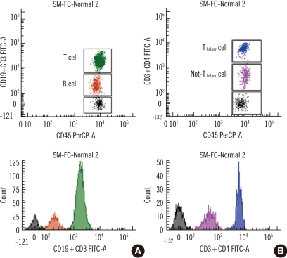
Fig. 1 Representative dot plots and histograms of single-color multitarget flow cytometry with CD19-FITCdim+CD3-FITCbright and with CD3-FITCdim+CD4-FITCbright. (A) B cells (CD19-FITCdim, brown) and T cells (CD3-FITCbright, green). (B) Non-Thelper cells (CD3-FITCdim, pink) and Thelper cells (CD3-FITCdim+CD4-FTTCbright, blue).Abbreviations: SM-FC, single-color multitarget flow cytometry; PerCP, peridinin chlorophyll protein complex; FITC, fluorescein α-isothyocyanate.
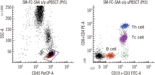
Fig. 2 Representative dot plots of single-color multitarget flow cytometry with CD19-FITCdim+CD3-FITCbright and CD8-PEdim+CD4-PEbright. B cells (CD19-FITCdim, brown), Tcytotoxic cells (CD3-FITCbright+CD8-PEdim, pink), and Thelper cells (CD3-FITCbright+CD4-PEbright, blue) in a right dot plot.Abbreviations: SM-FC-SAA s/p uPBSCT, single-color multitarget flow cytometry-severe aplastic anemia after unrelated peripheral blood stem cell transplantation; SSC-A, side scatter; PerCP, peridinin chlorophyll protein complex; PE, phycoerythrin; FITC, fluorescein α-isothyocyanate.
Reference
-
1. Hengel RL, Nicholson JK. An update on the use of flow cytometry in HIV infection and AIDS. Clin Lab Med. 2001; 21:841–856. PMID: 11770291.2. Illoh OC. Current applications of flow cytometry in the diagnosis of primary immunodeficiency diseases. Arch Pathol Lab Med. 2004; 128:23–31. PMID: 14692816.
Article3. Mandy FF. Twenty-five years of clinical flow cytometry: AIDS accelerated global instrument distribution. Cytometry A. 2004; 58:55–56. PMID: 14994221.
Article4. Braylan RC. Impact of flow cytometry on the diagnosis and characterization of lymphomas, chronic lymphoproliferative disorders and plasma cell neoplasias. Cytometry A. 2004; 58:57–61. PMID: 14994222.
Article5. Orfao A, Ortuño F, de Santiago M, Lopez A, San Miguel J. Immunophenotyping of acute leukemias and myelodysplastic syndromes. Cytometry A. 2004; 58:62–71. PMID: 14994223.
Article6. Keeney M, Gratama JW, Sutherland DR. Critical role of flow cytometry in evaluating peripheral blood hematopoietic stem cell grafts. Cytometry A. 2004; 58:72–75. PMID: 14994224.
Article7. Bagwell CB. DNA histogram analysis for node-negative breast cancer. Cytometry A. 2004; 58:76–78. PMID: 14994225.
Article8. Maecker HT, Maino VC, editors. Manual of Clinical Laboratory Immunology. 2002. 6th ed. Washington, DC: ASM Press;p. 338–346.9. Vermes I, Haanen C, Reutelingsperger C. Flow cytometry of apoptotic cell death. J Immunol Methods. 2000; 243:167–190. PMID: 10986414.
Article10. Lehmann AK, Sornes S, Halstensen A. Phagocytosis: measurement by flow cytometry. J Immunol Methods. 2000; 243:229–242. PMID: 10986417.
Article11. Krutzik PO, Irish JM, Nolan GP, Perez OD. Analysis of protein phosphorylation and cellular signaling events by flow cytometry: techniques and clinical applications. Clin Immunol. 2004; 110:206–221. PMID: 15047199.
Article12. Pala P, Hussell T, Openshaw PJ. Flow cytometric measurement of intracellular cytokines. J Immunol Methods. 2000; 243:107–124. PMID: 10986410.
Article13. Pozarowski P, Darzynkiewicz Z. Analysis of cell cycle by flow cytometry. Methods Mol Biol. 2004; 281:301–311. PMID: 15220539.
Article14. Gratama JW, Kraan J, Keeney M, Granger V, Barnett D. Reduction of variation in T-cell subset enumeration among 55 laboratories using single-platform, three or four-color flow cytometry based on CD45 and SSC-based gating of lymphocytes. Cytometry. 2002; 50:92–101. PMID: 12116351.
Article15. Alamo AL, Melnick SJ. Clinical application of four and five-color flow cytometry lymphocyte subset immunophenotyping. Cytometry. 2000; 42:363–370. PMID: 11135290.
Article16. Chng WJ, Tan GB, Kuperan P. Establishment of adult peripheral blood lymphocyte subset reference range for an Asian population by single-platform flow cytometry: influence of age, sex, and race and comparison with other published studies. Clin Diagn Lab Immunol. 2004; 11:168–173. PMID: 14715565.
Article17. Autissier P, Soulas C, Burdo TH, Williams KC. Evaluation of a 12-color flow cytometry panel to study lymphocyte, monocyte, and dendritic cell subsets in humans. Cytometry A. 2010; 77:410–419. PMID: 20099249.
Article18. Colombo F, Cattaneo A, Lopa R, Portararo P, Rebulla P, Porretti L. Evaluation of a multicolor, single-tube technique to enumerate lymphocyte subpopulations. Clin Vaccine Immunol. 2008; 15:1124–1127. PMID: 18448621.
Article19. Lambert C, Cristina I, Christian G. Enumeration of peripheral lymphocyte subsets using 6 vs. 4 color staining: a clinical evaluation of a new flowcytometer. Cytometry B Clin Cytom. 2006; 70:29–38. PMID: 16353133.
Article20. Ashman M, Sachdeva N, Davila L, Scott G, Mitchell C, Cintron L, et al. Influence of 4- and 6-color flow cytometers and acquisition/analysis softwares on the determination of lymphocyte subsets in HIV infection. Cytometry B Clin Cytom. 2007; 72:380–386. PMID: 17226862.
Article21. Szczepański T, van der Velden VH, van Dongen JJ. Flow-cytometric immunophenotyping of normal and malignant lymphocytes. Clin Chem Lab Med. 2006; 44:775–796. PMID: 16776621.
Article22. Roussel M, Benard C, Ly-Sunnaram B, Fest T. Refining the white blood cell differential: the first flow cytometry routine application. Cytometry A. 2010; 77:552–563. PMID: 20506466.
Article23. Cherian S, Levin G, Lo WY, Mauck M, Kuhn D, Lee C, et al. Evaluation of an 8-color flow cytometric reference method for white blood cell differential enumeration. Cytometry B Clin Cytom. 2010; 78:319–328. PMID: 20533390.
Article24. Björnsson S, Wahlström S, Norström E, Bernevi I, O'Neill U, Johansson E, et al. Total nucleated cell differential for blood and bone marrow using a single tube in a five-color flow cytometer. Cytometry B Clin Cytom. 2008; 74:91–103. PMID: 18061952.
Article25. Faucher JL, Lacronique-Gazaille C, Frébet E, Trimoreau F, Donnard M, Bordessoule D, et al. "6 markers/5 colors" extended white blood cell differential by flow cytometry. Cytometry A. 2007; 71:934–944. PMID: 17879238.
Article26. Swerdlow SH, Campo E, editors. WHO classification tumours of haematopoietic and lymphoid tissues. 2008. 4th ed. Lyon: IARC press;p. 109–320.
- Full Text Links
-
- Actions
-
Cited
- CITED
-
- Close
- Share
-
- Similar articles
-
- Production of the Monoclonal Antibodies against Bartonella henselae Isolated from a Korean Patient
- Multiparameter Flow Cytometry: Advances in High Resolution Analysis
- The Study of The Immunoassay by the Monoclonal Antibody: Flow Cytometry for Cell Surface Phenotyping
- Establishment of Reference Values for Platelet Activation Markers by Flow Cytometry
- Generation and Characterization of Anti - Human CTLA - 4 Monoclonal Antibodies
