Suture Granuloma Mimicking Recurrent Thyroid Carcinoma on Ultrasonography
- Affiliations
-
- 1Department of Diagnostic Radiology, Yonsei University College of Medicine, Seoul, Korea. ekkim@yumc.yonsei.ac.kr
- 2Department of Pathology, Yonsei University College of Medicine, Seoul, Korea.
- KMID: 1381259
- DOI: http://doi.org/10.3349/ymj.2006.47.5.748
Abstract
- Although high resolution ultrasonography (US) is helpful in the differentiation of suture granulomas from recurrent thyroid cancer in most cases, a definite diagnosis cannot always be made. We report a case that mimicked recurrent thyroid cancer on US and 2-[fluorine-18] fluoro-2-deoxy-D-glucose (FDG) positron emission tomography (PET), but diagnosis of a suture granuloma was confirmed by a US-guided fine needle aspiration biopsy (FNAB). In order to avoid unnecessary operations, the differential diagnosis between postoperative suture granulomas and recurrent cancer is important.
Keyword
MeSH Terms
-
Thyroidectomy/*adverse effects
Thyroid Neoplasms/diagnosis/pathology/ultrasonography
Sutures/*adverse effects
Positron-Emission Tomography
Neoplasm Recurrence, Local/diagnosis/pathology/ultrasonography
Humans
Granuloma, Foreign-Body/*diagnosis/etiology/ultrasonography
Female
Diagnosis, Differential
Biopsy, Fine-Needle
Adult
Figure
-

Fig. 1 Longitudinal US shows a 1.4 × 1.5 cm-sized ill-defined hypoechic nodule with microcalcification at the right thyroid gland. It was confirmed to be a papillary carcinoma by surgery.

Fig. 2 A, B. Transverse (A) and longitudinal (B) US shows an irregular shaped hypoechic nodule (arrows) in the right thyroidectomy bed.
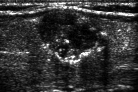
Fig. 3 A, B. Coronal (A) and Axial view (B). A few foci of mildly increased FDG uptake are seen in the right thyroidectomy area.
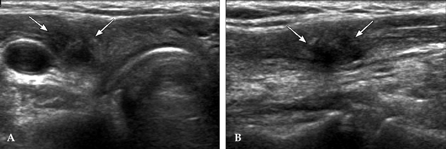
Fig. 4 (A) A multinucleated giant cell engulfs several acute inflammatory cells in the background of the fibrohistiocytic and neutrophilic cells (Papanicolaou × 400). (B) A large central irregular foreign material is surrounded by granulomatous inflammatory aggregates in the background of variable inflammatory cells and frequent scattered giant cells (Diff-Quick stain × 100).
Cited by 3 articles
-
Postoperative Surveillance of Thyroid Cancer: in View of US
Jin Young Kwak
J Korean Thyroid Assoc. 2012;5(1):15-19. doi: 10.11106/jkta.2012.5.1.15.Retained Drainage Tube Fragment after Thyroidectomy Presenting as a Palpable Neck Mass: a Case Report
Jung Han Yoon, Sin Jae Kang, Young Jae Ryu, Jin Seong Cho, Hyo Soon Lim, Ji Shin Lee, Min Ho Park
J Endocr Surg. 2017;17(4):149-152. doi: 10.16956/jes.2017.17.4.149.Postoperative Surveillance of Thyroid Cancer: In View of a Radiologist
Jin Young Kwak
J Korean Thyroid Assoc. 2015;8(1):8-13. doi: 10.11106/cet.2015.8.1.8.
Reference
-
1. Hegedüs L. Thyroid ultrasound. Endocrinol Metab Clin North Am. 2001. 30:339–360.2. Titton RL, Gervais DA, Boland GW, Maher MM, Mueller PR. Sonography and sonographically guided fine-needle aspiration biopsy of the thyroid gland: indications and techniques, pearls and pitfalls. AJR Am J Roentgenol. 2003. 181:267–271.3. Rettenbacher T, Macheiner P, Hollerweger A, Gritzmann N, Weismann C, Todoroff B. Suture granulomas: sonography enables a correct preoperative diagnosis. Ultrasound Med Biol. 2001. 27:343–350.4. Postlethwait RW, Willigan DA, Ulin AW. Human tissue reaction to sutures. Ann Surg. 1975. 181:144–150.5. Nagar H, Kessler A, Graif M. The role of ultrasound in the diagnosis of stitch granulomas following paediatric herniotomy. Pediatr Radiol. 1999. 29:803–806.6. Eldridge PR, Wheeler MH. Stitch granulomata after thyroid surgery. Br J Surg. 1987. 74:62.7. Khan N, Oriuchi N, Higuchi T, Zhang H, Endo K. PET in the follow-up of differentiated thyroid cancer. Br J Radiol. 2003. 76:690–695.8. Liu SH, Chang JT, Ng SH, Chan SC, Yen TC. False positive fluorine-18 fluorodeoxy-D-glucose positron emission tomography finding caused by osteoradionecrosis in a nasopharyngeal carcinoma patient. Br J Radiol. 2004. 77:257–260.9. Holder WD Jr, White RL Jr, Zuger JH, Easton EJ Jr, Greene FL. Effectiveness of positron emission tomography for the detection of melanoma metastases. Ann Surg. 1998. 227:764–771.
- Full Text Links
-
- Actions
-
Cited
- CITED
-
- Close
- Share
-
- Similar articles
-
- A Case of Rectal Suture Granuloma that was Suspected to be a Recurrent Rectal Carcinoma
- A Case of Zenker's Diverticulum Mimicking a Right Side Thyroid Nodule
- Knot-and-ear sign: a pathognomonic ultrasonographic feature of suture granuloma after thyroid surgery
- Branchial Cleft Cyst Mimicking Malignant Thyroid Neoplasm Concurrent with Medullary and Papillary Thyroid Carcinoma: a Case Report
- Ultrasonographic imaging of papillary thyroid carcinoma variants
