Korean J Ophthalmol.
2012 Jun;26(3):182-188. 10.3341/kjo.2012.26.3.182.
Low Limit for Effective Signal Strength in the Stratus OCT in Imperative Low Signal Strength Cases
- Affiliations
-
- 1Department of Ophthalmology, Kangbuk Samsung Hospital, Sungkyunkwan University School of Medicine, Seoul, Korea. kjoonmo1@gmail.com
- 2Department of Applied Statistics, Yonsei University, Seoul, Korea.
- 3Department of Ophthalmology, Seoul National University College of Medicine, Seoul, Korea.
- 4Department of Ophthalmology, Seoul National University Bundang Hospital, Seongnam, Korea.
- KMID: 1376123
- DOI: http://doi.org/10.3341/kjo.2012.26.3.182
Abstract
- PURPOSE
To determine the lowest limit of signal strength that is still effective for accurate analysis of optic coherence tomography (OCT) values, we investigated the reproducibility of OCT scans by signal strength (SS).
METHODS
A total of 668 subjects were scanned for measurements of retinal nerve fiber layer (RNFL) thickness using the Stratus OCT twice on the same day. The variability of overall RNFL thickness parameters obtained at different SS was analyzed and compared by repeated-measures of ANOVA and Spearman's correlation coefficient. Values of the intraclass correlation coefficient (ICC) and variability (standard deviation) of RNFL thickness were obtained. The false positive ratio was analyzed.
RESULTS
When SS was 3, the variability of RNFL thickness was significantly different (low ICC, high variability) in comparison to when SS was 4 or greater. Significant negative correlations were observed between variability in RNFL thickness and signal strength. The difference of variability of average RNFL thickness between SS 4 (4.94 microm) and SS 6 (4.41 microm) was 0.53 microm.
CONCLUSIONS
Clinically, the difference of variability of average RNFL thickness between SS 4 and SS 6 was quite small. High SS is important, however, when signal strength is low due to uncorrectable factors in patients in need of OCT for glaucoma and retinal disease. Our results suggest that SS 4 is the lowest acceptable limit of signal strength for obtaining reproducible scanning images.
Keyword
MeSH Terms
Figure
-
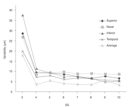
Fig. 1 The variability of retinal nerve fiber layer (RNFL) thickness by signal strength (SS). When SS was 3, the variability of RNFL thickness increased significantly, as opposed to when signal strength was 4 or greater.

Fig. 2 The box-plot diagram of correlations between the coefficient of variation (CV) in average retinal nerve fiber layer (RNFL) thickness, the RNFL thickness of four quadrants, the 12 o'clock hour (×) RNFL thickness, and signal strength. Significant negative correlations were observed between CV in RNFL thickness and signal strength (SS), indicating that decreased SS is associated with increased variability in RNFL thickness.
Reference
-
1. Larsson E, Eriksson U, Alm A. Retinal nerve fibre layer thickness in full-term children assessed with Heidelberg retinal tomography and optical coherence tomography: normal values and interocular asymmetry. Acta Ophthalmol. 2011. 89:151–158.2. Patel PJ, Chen FK, Ikeji F, Tufail A. Intersession repeatability of optical coherence tomography measures of retinal thickness in early age-related macular degeneration. Acta Ophthalmol. 2011. 89:229–234.3. Eriksson U, Alm A. Repeatability in and interchangeability between the macular and the fast macular thickness map protocols: a study on normal eyes with Stratus optical coherence tomography. Acta Ophthalmol. 2009. 87:725–730.4. Stein DM, Ishikawa H, Hariprasad R, et al. A new quality assessment parameter for optical coherence tomography. Br J Ophthalmol. 2006. 90:186–190.5. Ray R, Stinnett SS, Jaffe GJ. Evaluation of image artifact produced by optical coherence tomography of retinal pathology. Am J Ophthalmol. 2005. 139:18–29.6. El-Ashry M, Appaswamy S, Deokule S, Pagliarini S. The effect of phacoemulsification cataract surgery on the measurement of retinal nerve fiber layer thickness using optical coherence tomography. Curr Eye Res. 2006. 31:409–413.7. Savini G, Zanini M, Barboni P. Influence of pupil size and cataract on retinal nerve fiber layer thickness measurements by Stratus OCT. J Glaucoma. 2006. 15:336–340.8. Cheung CY, Leung CK, Lin D, et al. Relationship between retinal nerve fiber layer measurement and signal strength in optical coherence tomography. Ophthalmology. 2008. 115:1347–1351. 1351.e1–1351.e2.9. Youm DJ, Kim JM, Park KH, Choi CY. The effect of soft contact lenses during the measurement of retinal nerve fiber layer thickness using optical coherence tomography. Curr Eye Res. 2009. 34:78–83.10. Helb HM, Charbel Issa P, Fleckenstein M, et al. Clinical evaluation of simultaneous confocal scanning laser ophthalmoscopy imaging combined with high-resolution, spectral-domain optical coherence tomography. Acta Ophthalmol. 2010. 88:842–849.11. Li S, Wang X, Li S, et al. Evaluation of optic nerve head and retinal nerve fiber layer in early and advance glaucoma using frequency-domain optical coherence tomography. Graefes Arch Clin Exp Ophthalmol. 2010. 248:429–434.12. Gordon MO, Beiser JA, Brandt JD, et al. The Ocular Hypertension Treatment Study: baseline factors that predict the onset of primary open-angle glaucoma. Arch Ophthalmol. 2002. 120:714–720.13. Katsanos A, Labiris G, Fanariotis M, et al. The relationship between Rarebit perimetry and OCT-derived retinal nerve fibre layer thickness in glaucoma. Acta Ophthalmol. 2008. 86:871–876.14. Iester M, Garway-Heath D, Lemij HG, et al. Optic nerve head and retinal nerve fibre analysis. 2005. Savona: Editrice Dogma;121–149.15. Hougaard JL, Heijl A, Bengtsson B. Glaucomatous retinal nerve fibre layer defects may be identified in Stratus OCT images classified as normal. Acta Ophthalmol. 2008. 86:569–575.16. Teikari JM, O'Donnell J. Epidemiologic data on adult glaucomas. Data from the hospital discharge registry and the registry of right to free medication. Acta Ophthalmol (Copenh). 1989. 67:184–191.17. Tsai JC. Optical coherence tomography measurement of retinal nerve fiber layer after acute primary angle closure with normal visual field. Am J Ophthalmol. 2006. 141:970–972.18. DeLeon Ortega JE, Sakata LM, Kakati B, et al. Effect of glaucomatous damage on repeatability of confocal scanning laser ophthalmoscope, scanning laser polarimetry, and optical coherence tomography. Invest Ophthalmol Vis Sci. 2007. 48:1156–1163.19. Huynh SC, Wang XY, Rochtchina E, Mitchell P. Peripapillary retinal nerve fiber layer thickness in a population of 6-year-old children: findings by optical coherence tomography. Ophthalmology. 2006. 113:1583–1592.20. Repka MX, Kraker RT, Tamkins SM, et al. Retinal nerve fiber layer thickness in amblyopic eyes. Am J Ophthalmol. 2009. 148:143–147.21. Salchow DJ, Oleynikov YS, Chiang MF, et al. Retinal nerve fiber layer thickness in normal children measured with optical coherence tomography. Ophthalmology. 2006. 113:786–791.22. Yu S, Tanabe T, Hangai M, et al. Scanning laser polarimetry with variable corneal compensation and optical coherence tomography in tilted disk. Am J Ophthalmol. 2006. 142:475–482.23. Huang ML, Chen HY, Lin JC. Rule extraction for glaucoma detection with summary data from Stratus OCT. Invest Ophthalmol Vis Sci. 2007. 48:244–250.24. Fisher JB, Jacobs DA, Markowitz CE, et al. Relation of visual function to retinal nerve fiber layer thickness in multiple sclerosis. Ophthalmology. 2006. 113:324–332.25. Shah NN, Bowd C, Medeiros FA, et al. Combining structural and functional testing for detection of glaucoma. Ophthalmology. 2006. 113:1593–1602.26. Kim TW, Zangwill LM, Bowd C, et al. Retinal nerve fiber layer damage as assessed by optical coherence tomography in eyes with a visual field defect detected by frequency doubling technology perimetry but not by standard automated perimetry. Ophthalmology. 2007. 114:1053–1057.27. Wu Z, Huang J, Dustin L, Sadda SR. Signal strength is an important determinant of accuracy of nerve fiber layer thickness measurement by optical coherence tomography. J Glaucoma. 2009. 18:213–216.28. Wu Z, Vazeen M, Varma R, et al. Factors associated with variability in retinal nerve fiber layer thickness measurements obtained by optical coherence tomography. Ophthalmology. 2007. 114:1505–1512.
- Full Text Links
-
- Actions
-
Cited
- CITED
-
- Close
- Share
-
- Similar articles
-
- The Effect of Machine Aging on the Measurements of Optical Coherency Tomography
- Analysis of Factors Associated with Variability in Measures Obtained by Spectral Domain Optical Coherence Tomography
- Comparison of Retinal Nerve Fiber Layer Thickness Measured by Spectral-Domain and Time-Domain Optical Coherence Tomography
- Comparison of Time Domain OCT and Spectrum Domain OCT for Retinal Nerve Fiber Layer Assessment
- Usefulness of Table Parameters of Stratus OCT in Detection of Localized Retinal Nerve Fiber Layer Defects
