J Korean Rheum Assoc.
2008 Jun;15(2):118-122. 10.4078/jkra.2008.15.2.118.
Increased Interleukin-17 Expression in Patients with Idiopathic Inflammatory Myopathies
- Affiliations
-
- 1Department of Internal Medicine, College of Medicine, Pusan National University, Busan, Korea. ksimd@pusan.ac.kr
- 2Department of Neurology, College of Medicine, Pusan National University, Busan, Korea.
- 3Research Institute, College of Medicine, Pusan National University, Busan, Korea.
- 4Department of Internal Medicine, Busan St. Mary's Medical Center, Busan, Korea.
- KMID: 1270227
- DOI: http://doi.org/10.4078/jkra.2008.15.2.118
Abstract
- OBJECTIVE: Idiopathic inflammatory myopathies (IIMs) are systemic autoimmune diseases characterized by infiltration of T lymphocytes, monocytes, and macrophages in muscle tissues. Interleukin-17 (IL-17), a Th17 cytokine, has potent pro-inflammatory actions and plays a role in autoimmune diseases. We investigated the expression of IL-17 in muscle tissues of patients with IIMs. METHODS: We measured the IL-17 mRNA level of muscle tissues from 14 patients with IIMs (9 patients with dermatomyositis and 5 patients with polymyositis) by real-time RT-PCR and compared with controls. We also performed an immunohistochemical stain to detect IL-17 expression. RESULTS: The expressions of IL-17 were significantly enhanced in IIMs than controls. In immunohistochemistry, IL-17 was expressed in perimysial, endomysial and perivascular infiltrating inflammatory cells. CONCLUSION: These results suggest that IL-17 plays a role in the immunopathogenesis of IIMs.
Figure
-
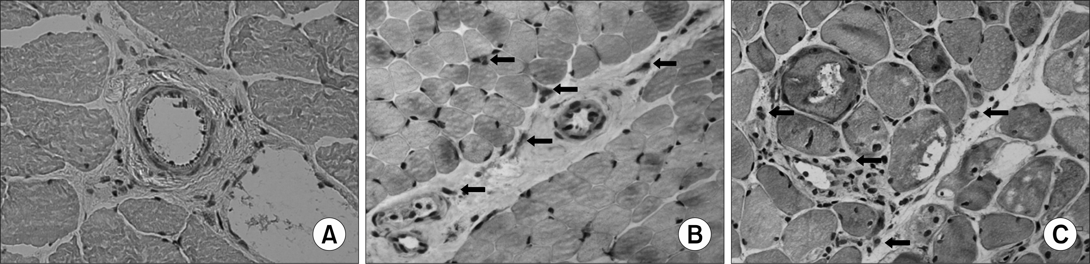
Fig. 1. Immunohistochemistry of IL-17 in muscle tissues from patients with IIMs (B, C) and control (A). In DM (B) and PM (C), IL-17 was expressed in perimysial, endomysialand perivascular infiltrating inflammatory cells (arrow). Muscle tissue from control was negative (A). Magnification, ×200. IIMs: idiopathic inflammatory myopathies, DM: dermatomyositis, PM: polymyositis.
Reference
-
References
1. Briani C, Doria A, Sarzi-Puttini P, Dalakas MC. Update on idiopathic inflammatory myopathies. Autoimmunity. 2006; 39:161–70.
Article2. Wiendl H, Hoflfeld R, Kieseier BC. Immunobiology of muscle: advances in understanding an immunological microenvironment. Trend Immunol. 2005; 26:373–80.
Article3. Salomonsson S, Lundberg I. Cytokines in idiopathic inflammatory myopathies. Autoimmunity. 2006; 39:177–90.
Article4. McFarland HF, Martin R. Multiple sclerosis: a complicated picture of autoimmunity. Nat Immunol. 2007; 8:913–9.
Article5. Toh ML, Miossec P. The role of T cells in rheumatoid arthritis: new subsets and new targets. Curr Opin Rheumatol. 2007; 19:284–8.
Article6. Chevrel G, Page G, Granet C, Streichenberger N, Varennes A, Miossec P. Interleukin-17 increases the effects of IL-1 beta on muscle cells: arguments for the role of T cells in the pathogenesis of myositis. J Neuroimmunol. 2003; 137:125–33.7. Page G, Chevrel G, Miossec P. Anatomic localization of immature and mature dendritic cell subsets in dermatomyositis and polymyositis. Arthritis Rheum. 2004; 50:199–208.8. Baird GS, Montine TJ. Multiplex immunoassay anlysis of cytokines in idiopathic inflammatory myo-pahty. Arch Pathol Lab Med. 2008; 132:232–8.9. Bohan A, Peter JB. Polymyositis and dermatomyositis (first of two parts). N Engl J Med. 1975; 292:403–7.10. Bohan A, Peter JB. Polymyositis and dermatomyositis (second of two parts). N Engl J Med. 1975; 292:403–7.
- Full Text Links
-
- Actions
-
Cited
- CITED
-
- Close
- Share
-
- Similar articles
-
- The Expression of Toll-like Receptors in Idiopathic Inflammatory Myopathies
- Immunohistochemical expression in idiopathic inflammatory myopathies at a single center in Vietnam
- Extensive inflammatory reaction in facioscapulohumeral muscular dystrophy
- Effects of Dexamethasone on expressions of IFN-gamma and IL-4 by PBMCs in response to IL-12
- The Laboratory Test for the Diagnosis of Idiopathic Inflammatory Myopathies
