US-Guided Vacuum-Assisted Biopsy of Microcalcifications in Breast Lesions and Long-Term Follow-Up Results
- Affiliations
-
- 1Department of Radiology and Institution of Radiological Science, Severance Hospital, Yonsei University College of Medicine, Seoul, Korea. mines@yuhs.ac
- 2Department of Diagnostic Radiology, Yongdong Severance Hospital, Yonsei University College of Medicine, Seoul, Korea.
- KMID: 1118871
- DOI: http://doi.org/10.3348/kjr.2008.9.6.503
Abstract
OBJECTIVE
To evaluate the diagnostic accuracy of the use of an ultrasonography (US)-guided vacuum-assisted biopsy for microcalcifications of breast lesions and to evaluate the efficacy of the use of US-guided vacuum-assisted biopsy with long-term follow-up results. MATERIALS AND METHODS: US-guided vacuum-assisted biopsy cases of breast lesions that were performed between 2002 and 2006 for microcalcifications were retrospectively reviewed. A total of 62 breast lesions were identified where further pathological confirmation was obtained or where at least two years of mammography follow-up was obtained. These lesions were divided into the benign and malignant lesions (benign and malignant group) and were divided into underestimated group and not-underestimated lesions (underestimated and not-underestimated group) according to the diagnosis after a vacuum-assisted biopsy. The total number of specimens that contained microcalcifications was analyzed and the total number of microcalcification flecks as depicted on specimen mammography was analyzed to determine if there was any statistical difference between the groups. RESULTS: There were no false negative cases after more than two years of follow-up. Twenty-nine lesions were diagnosed as malignant (two invasive carcinomas and 27 carcinoma in situ lesions). Two of the 27 carcinoma in situ lesions were upgraded to invasive cancers after surgery. Among three patients diagnosed with atypical ductal hyperplasia, the diagnosis was upgraded to a ductal carcinoma in situ after surgery in one patient. There was no statistically significant difference in the number of specimens with microcalcifications and the total number of microcalcification flecks between the benign group and malignant group of patients and between the underestimated group and not-underestimated group of patients. CONCLUSION: US-guided vacuum-assisted biopsy can be an effective alternative to stereotactic-guided vacuum-assisted biopsy in cases where microcalcifications are visible with the use of high-resolution US.
Keyword
MeSH Terms
Figure
-
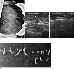
Fig. 1 44-year-old female who underwent mammography for routine breast screening. A. Magnification view of left breast reveals suspicious clustered and pleomorphic microcalcification (white arrow). B. Careful US study of left breast shows microcalcification of suspicion (white arrows) in left upper central aspect. C. Patient underwent 11 gauge US-guided vacuam-assisted biopsy targeted at microcalcification. D. Specimen mammography shows that aimed microcalcification is retrieved (white arrows). Patient was diagnosed with ductal carcinoma in situ and underwent further partial mastectomy, which also revealed presence of ductal carcinoma in situ.
Cited by 1 articles
-
Clinical Experience of Ultrasound-Guided, Vacuum-Assisted Breast Biopsy for Mammographic Microcalcifications: Combination with Wire Localization
SeungSang Ko, Man Sik Shin, Ki Won Chun, Kang Young Rhee, Heeboong Park
J Surg Ultrasound. 2018;5(2):53-60. doi: 10.46268/jsu.2018.5.2.53.
Reference
-
1. Parker SH, Burbank F, Jackman RJ, Aucreman CJ, Cardenosa G, Cink TM, et al. Percutaneous large-core breast biopsy: a multi-institutional study. Radiology. 1994. 193:359–364.2. Meyer JE, Smith DN, Lester SC, Kaelin C, DiPiro PJ, Denison CM, et al. Large-core needle biopsy of nonpalpable breast lesions. JAMA. 1999. 281:1638–1641.3. Brenner RJ, Bassett LW, Fajardo LL, Dershaw DD, Evans WP 3rd, Hunt R, et al. Stereotactic core-needle breast biopsy: a multi-institutional prospective trial. Radiology. 2001. 218:866–872.4. Philpotts LE, Hooley RJ, Lee CH. Comparison of automated versus vacuum-assisted biopsy methods for sonographically guided core biopsy of the breast. AJR Am J Roentgenol. 2003. 180:347–351.5. Liberman L. Percutaneous image-guided core breast biopsy. Radiol Clin North Am. 2002. 40:483–500.6. DeAngelis GA, Moran RE, Fajardo LL, Mugler JP, Christopher JM, Harvey JA. MRI-guided needle localization: technique. Semin Ultrasound CT MR. 2000. 21:337–350.7. Youk JH, Kim EK, Kim MJ, Lee JY, Oh KK. Missed breast cancers at US-guided core needle biopsy: How to reduce them. Radiographics. 2007. 27:79–94.8. American College of Radiology. Breast imaging reporting and data system (BI-RADS). 2003. 4th ed. Reston: Va: America College of Radiology.9. Cady B, Stone MD, Schuler JG, Thakur R, Wanner MA, Lavin PT. The new era in breast cancer. Invasion, size, and nodal involvement dramatically decreasing as a result of mammographic screening. Arch Surg. 1996. 131:301–308.10. Holland R, Hendriks JH, Vebeek AL, Mravunac M, Schuurmans Stekhoven JH. Extent, distribution, and mammographic/histological correlations of breast ductal carcinoma in situ. Lancet. 1990. 335:519–522.11. Spencer NJ, Evans AJ, Galea M, Sibbering DM, Yeoman LJ, Pinder SE, et al. Pathological-radiological correlations in benign lesions excised during a breast screening programme. Clin Radiol. 1994. 49:853–856.12. Jackman RJ, Marzoni FA Jr. Needle-localized breast biopsy: why do we fail? Radiology. 1997. 204:677–684.13. Head JF, Haynes AE, Elliott MC, Elliott RL. Stereotaxic localization and core needle biopsy of nonpalpable breast lesions: two-year follow-up of a prospective study. Am Surg. 1996. 62:1018–1023.14. Elvecrog EL, Lechner MC, Nelson MT. Nonpalpable breast lesions: correlation of stereotaxic large-core needle biopsy and surgical biopsy results. Radiology. 1993. 188:453–455.15. Soo MS, Baker JA, Rosen EL. Sonographic detection and sonographically guided biopsy of breast microcalcifications. AJR Am J Roentgenol. 2003. 180:941–948.16. Cangiarella J, Waisman J, Symmans WF, Gross J, Cohen JM, Wu H, et al. Mammotome core biopsy for mammary microcalcification: analysis of 160 biopsies from 142 women with surgical and radiologic follow-up. Cancer. 2001. 91:173–177.17. Liberman L, Smolkin JH, Dershaw DD, Morris EA, Abramson AF, Rosen PP. Calcification retrieval at stereotactic, 11-gauge, directional, vacuum-assisted breast biopsy. Radiology. 1998. 208:251–260.18. Meyer JE, Smith DN, DiPiro PJ, Denison CM, Frenna TH, Harvey SC, et al. Stereotactic breast biopsy of clustered microcalcifications with a directional, vacuum-assisted device. Radiology. 1997. 204:575–576.19. Kettritz U, Rotter K, Schreer I, Murauer M, Schulz-Wendtland R, Peter D, et al. Stereotactic vacuum-assisted breast biopsy in 2874 patients: a multicenter study. Cancer. 2004. 100:245–251.20. Kim YM, Park HB, Ryu JW. Usefulness of ultrasound-guided mammotome biopsy for microcalcification. J Korean Radiol Soc. 2005. 53:129–135. [Korean].21. Jackman RJ, Burbank F, Parker SH, Evans WP 3rd, Lechner MC, Richardson TR, et al. Atypical ductal hyperplasia diagnosed at stereotactic breast biopsy: Improved reliability with 14-gauge, directional, vacuum-assisted biopsy. Radiology. 1997. 204:485–488.22. Liberman L, Cohen MA, Dershaw DD, Abramson AF, Hann LE, Rosen PP. Atypical ductal hyperplasia diagnosed at stereotaxic core biopsy of breast lesions: an indication for surgical biopsy. AJR Am J Roentgenol. 1995. 164:1111–1113.23. Bagnall MJ, Evans AJ, Wilson AR, Burrell H, Pinder SE, Ellis IO. When have mammographic calcifications been adequately sampled at needle core biopsy? Clin Radiol. 2000. 55:548–553.
- Full Text Links
-
- Actions
-
Cited
- CITED
-
- Close
- Share
-
- Similar articles
-
- Usefulness of Ultrasound-guided Mammotome Biopsy for Microcalcification
- Benign core biopsy of probably benign breast lesions 2 cm or larger: correlation with excisional biopsy and long-term follow-up
- Breast Microcalcifications: Diagnostic Outcomes According to Image-Guided Biopsy Method
- Usefulness of Ultrasound Guided Vacuum-Assisted Mammotome Biopsy for Breast Lesion
- Papillary Lesions of the Breast: Comparison of the US-guided 14-Gauge Automated Gun Method and the 11-Gauge Directional Vacuum-Assisted Biopsy Method
