Focal Hepatic Lesions: Contrast-Enhancement Patterns at Pulse-Inversion Harmonic US using a Microbubble Contrast Agent
- Affiliations
-
- 1Department of Radiology and Institute of Medical Science, Wonkwang University School of Medicine. yoonkh@wmc.wonkwang.ac.kr
- KMID: 1111251
- DOI: http://doi.org/10.3348/kjr.2003.4.4.224
Abstract
OBJECTIVE
To analyze the contrast-enhancement patterns obtained at pulseinversion harmonic imaging (PIHI) of focal hepatic lesions, and to thus determine tumor vascularity and the acoustic emission effect. MATERIALS AND METHODS: We reviewed pulse-inversion images in 90 consecutive patients with focal hepatic lesions, namely hepatocellular carcinoma (HCC) (n=43), metastases (n=30), and hemangioma (n=17). Vascular and delayed phase images were obtained immediately and five minutes following the injection of a microbubble contrast agent. Tumoral vascularity at vascular phase imaging and the acoustic emission effect at delayed phase imaging were each classified as one of four patterns. RESULTS: Vascular phase images depicted internal vessels in 93% of HCCs, marginal vessels in 83% of metastases, and peripheral nodular enhancement in 71% of hemangiomas. Delayed phase images showed inhomogeneous enhancement in 86% of HCCs; hypoechoic, decreased enhancement in 93% of metastases; and hypoechoic and reversed echogenicity in 65% of hemangiomas. Vascular and delayed phase enhancement patterns were associated with a specificity of 91% or greater, and 92% or greater, respectively, and with positive predictive values of 71% or greater, and 85% or greater, respectively. CONCLUSION: Contrast-enhancement patterns depicting tumoral vascularity and the acoustic emission effect at PIHI can help differentiate focal hepatic lesions.
Keyword
MeSH Terms
-
Adult
Aged
Carcinoma, Hepatocellular/blood supply/*ultrasonography
Colon/pathology
Contrast Media/*administration & dosage
Diagnosis, Differential
Female
Hemangioma/blood supply/*ultrasonography
Human
Image Enhancement/*methods
Liver/pathology/ultrasonography
Liver Neoplasms/blood supply/secondary/*ultrasonography
Lung/pathology
Male
*Microbubbles
Middle Aged
Pancreas/pathology
Polysaccharides/administration & dosage/diagnostic use
Reproducibility of Results
Retrospective Studies
Sensitivity and Specificity
Stomach/pathology
Support, Non-U.S. Gov't
Figure
-
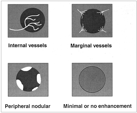
Fig. 1 The contrast-enhancement patterns observed at vascular phase pulse-inversion harmonic imaging of hepatic tumors.
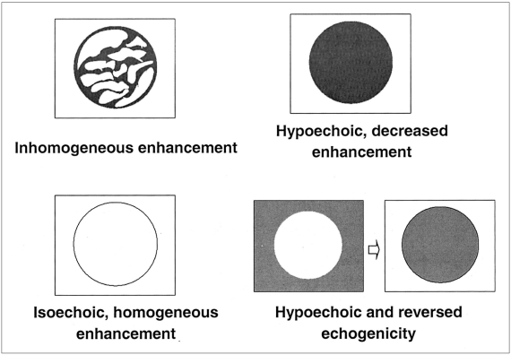
Fig. 2 The contrast-enhancement patterns seen at delayed phase pulse-inversion harmonic imaging of hepatic tumors.
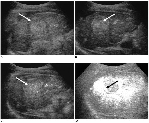
Fig. 3 Hepatocellular carcinoma showing the 'internal vessels' pattern during the vascular phase and 'inhomogeneous enhancement' during the delayed phase.A. Pulse-inversion harmonic US image before injection of the contrast agent depicts a hyperechoic mass (arrow).B, C. Vascular phase pulse-inversion harmonic US low-MI images obtained 15 sec (B) and 45 sec (C) after contrast injection depict the tumor's internal vessels (arrows).D. Delayed phase image (high MI) obtained five minutes after contrast injection demonstrates inhomogeneous enhancement (arrow) and the acoustic emission effect in hepatic parenchyma.
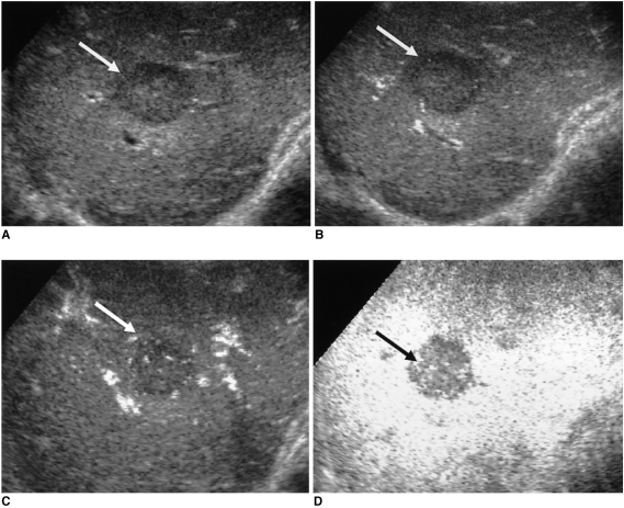
Fig. 4 Metastases from colon cancer showing 'marginal vessels' and 'hypoechoic, decreased enhancement'.A. Pulse-inversion harmonic US image obtained before contrast injection shows a hypoechoic mass (arrow).B, C. Vascular phase pulse-inversion harmonic US images (low MI) obtained 35 sec (B) and 54 sec (C) after contrast injection depict marginal tumor vessels (arrows) and some central vessels.D. Delayed phase image (high MI) shows a hypoechoic mass with decreased enhancement (arrow) compared to the acoustic emission effect of surrounding normal hepatic parenchyma.
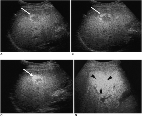
Fig. 5 Hemangioma demonstrating 'peripheral nodular enhancement' and 'isoechoic, homogeneous enhancement' patterns.A, B, C. Vascular phase pulse-inversion harmonic US images obtained 24 sec (A), 38 sec (B) and 56 sec (C) after contrast injection depict peripheral nodular enhancement with progressive centripetal fill-in (arrows).D. Delayed phase image (high MI) demonstrates isoechoic, homogeneous enhancement (arrowheads), which is the same appearance as normal hepatic parenchyma.
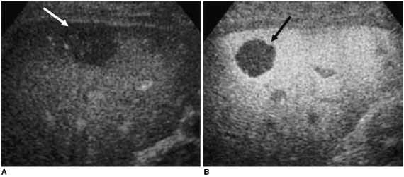
Fig. 6 Metastasis from stomach cancer, exhibiting the 'hypoechoic, decreased enhancement' pattern on delayed phase image.A. Pulse-inversion harmonic US image obtained prior to contrast injection depicts a hypoechoic nodule (arrow).B. Delayed phase image (high MI) shows a hypoechoic mass with decreased enhancement (arrow) compared to homogenous bright echogenic hepatic parenchyma.
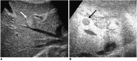
Fig. 7 Hemangioma demonstrating 'hypoechoic and reversed echogenicity' at the delayed phase.A. Conventional sonogram depicts a hyperechoic hepatic nodule (arrow).B. Delayed phase pulse-inversion harmonic image reveals a hypoechoic nodule with decreased enhancement compared to adjacent liver (arrow), and hypoechoic and reversed echogenicity at stimulated acoustic emission imaging.
Reference
-
1. Choi BI, Kim TK, Han JK, Chung JW, Park JH, Han MC. Power versus conventional color Doppler sonography: comparison in the depiction of vasculature in liver tumors. Radiology. 1996; 200:55–58. PMID: 8657945.
Article2. Lencioni R, Pinto F, Armillotta N, Bartolozzi C. Assessment of tumor vascularity in hepatocellular carcinoma: comparison of power Doppler US and color Doppler US. Radiology. 1996; 201:353–358. PMID: 8888222.
Article3. Burns PN. Harmonic imaging with ultrasound contrast agents. Clin Radiol. 1996; 51:50–55. PMID: 8605774.4. Goldberg BB, Liu J, Burns PN, Merton DA, Forsberg F. Galactose-based intravenous sonographic contrast agent: experimental studies. J Ultrasound Med. 1993; 12:463–470. PMID: 8411330.
Article5. Cosgrove D. Why do we need contrast agents for ultrasound? Clin Radiol. 1996; 51:1–4. PMID: 8605763.6. Schineider M, Broillet A, Bussat P, et al. Gray-scale liver enhancement in VX2 tumor-bearing rabbits using BR14, a new ultrasonographic contrast agent. Invest Radiol. 1997; 32:410–417. PMID: 9228607.7. Forsberg F, Goldberg BB, Liu J, Merton DA, Rawool NM, Shi WT. Tissue-specific US contrast agent for evaluation of hepatic and splenic parenchyma. Radiology. 1999; 210:125–132. PMID: 9885597.
Article8. Hauff PD, Fritzsch TD, Reinhardt M, et al. Delineation of experimental liver tumors in rabbits by a new ultrasound contrast agent and stimulated acoustic emission. Invest Radiol. 1997; 32:94–99. PMID: 9039581.
Article9. Kono Y, Moriyasu F, Nada T, et al. Gray scale second harmonic imaging of the liver: a preliminary animal study. Ultrasound Med Biol. 1997; 23:719–726. PMID: 9253819.
Article10. Burns PN, Wilson SR, Simpson DH. Pulse inversion imaging of liver blood flow: improved method for characterizing focal masses with microbubble contrast. Invest Radiol. 2000; 35:58–71. PMID: 10639037.11. Wilson SR, Burns PN, Muradali D, Wilson JA, Lai X. Harmonic hepatic US with microbubble contrast agent: initial experience showing improved characterization of hemangioma, hepatocellular carcinoma, and metastasis. Radiology. 2000; 215:153–161. PMID: 10751481.
Article12. Blomley MJK, Albrecht T, Cosgrove DO, et al. Improved imaging of liver metastases with stimulated acoustic emission in the late phase of enhancement with the US contrast agent SH U 508A: early experience. Radiology. 1999; 210:409–416. PMID: 10207423.
Article13. Dalla-Palma L, Bertolotto M, Quaia E, Locatelli M. Detection of liver metastases with pulse-inversion harmonic imaging: preliminary results. Eur Radiol. 1999; 9:S382–S387. PMID: 10602934.14. Kim TK, Choi BI, Han JK, Hong HS, Park SH, Moon SG. Hepatic tumors: contrast agent-enhancement patterns with pulse-inversion harmonic US. Radiology. 2000; 216:411–417. PMID: 10924562.
Article15. Jang HJ, Lim HK, Lee WJ, et al. Focal hepatic lesions: evaluation with contrast-enhanced gray-scale harmonic US. Korean J Radiol. 2003; 4:91–100. PMID: 12845304.
Article16. Dill-Macky MJ, Burns PN, Khalili K, Wilson SR. Focal hepatic masses: enhancement patterns with SH U 508A and pulse-inversion US. Radiology. 2002; 222:95–102. PMID: 11756711.
Article17. Albrecht T, Hoffmann CW, Schmitz S, et al. Phase-inversion sonography during the liver-specific late phase of contrast enhancement: improved detection of liver metastases. AJR Am J Roentgenol. 2001; 176:1191–1198. PMID: 11312180.18. Blomley MJK, Albrecht T, Cosgrove DO, et al. Stimulated acoustic emission to image a late liver and spleen-specific phase of Levovist® in normal volunteers and patients with and without liver disease. Ultrasound Med Biol. 1999; 25:1341–1352. PMID: 10626621.
Article19. Ko CJ, Yoon KH. Gray-scale stimulated acoustic emission: differential diagnosis between hepatocellular carcinoma and metastatic adenocarcinoma. J Korean Radiol Soc. 2001; 44:63–68.
- Full Text Links
-
- Actions
-
Cited
- CITED
-
- Close
- Share
-
- Similar articles
-
- The Detectability of Hepatic Metastases in Candidates of Radiofrequency Ablation: Comparison for Helical CT Scanning and Late-Phase Pulse-Inversion Harmonic Imaging
- Detection of Hepatic VX2 Tumors in Rabbits: Comparison of Conventional US and Phase-Inversion Harmonic US During the Liver-Specific Late Phase of Contrast Enhancement
- Hepatic Hemangiomas: Spectrum of US Appearances on Gray-scale, Power Doppler, and Contrast-Enhanced US
- Focal Hepatic Lesions: Evaluation with Contrast-Enhanced Gray-Scale Harmonic US
- The Study of Renal Perfusion Image in Rabbit by Harmonic Ultrasound with Microbble Contrast Agent in Comparison with 99mTc-DTPA: Focusing on US Scan Technique and Concentration of Contrast Agent
