J Vet Sci.
2010 Jun;11(2):155-159. 10.4142/jvs.2010.11.2.155.
Spectral Doppler ultrasound in the major arteries of normal conscious immature micropigs
- Affiliations
-
- 1Department of Veterinary Radiology, College of Veterinary Medicine, Seoul National University, Seoul 151-742, Korea. mcchoi@snu.ac.kr
- KMID: 1110863
- DOI: http://doi.org/10.4142/jvs.2010.11.2.155
Abstract
- Spectral waveform analysis of blood flow velocity in the major arteries of six healthy, conscious immature micropigs was determined using Doppler ultrasonography. Doppler spectral tracings were recorded from the external iliac artery, femoral artery, and renal arcuate artery. Tracings were also taken from three parts of the common carotid artery and two parts of the abdominal aorta. Spectral Doppler parameters included peak systolic velocity, early diastolic velocity, peak systolic velocity-to-end diastolic velocity ratio, resistive index, and pulsatility index. In addition, the diameter of major arteries and indirect blood pressure were measured. These results from spectral Doppler analysis in major arteries may be useful as reference ranges in the future studies of vascular hemodynamics in immature micropigs.
MeSH Terms
Figure
-
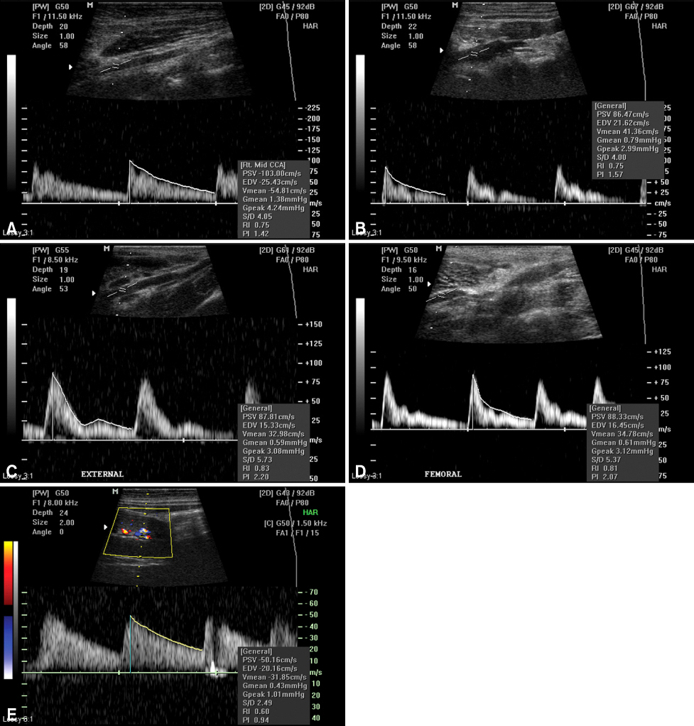
Fig. 1 Representative pulsed wave Doppler spectral waveform pattern for the common carotid artery (A), abdominal aorta (B), external iliac artery (C), femoral artery (D), and renal arcuate artery (E). The scale to the right of the image is in increments of 10 cm/sec.
Reference
-
1. Allan PL, Dubbins PA, Pozniak MA, McDicken WN. Clinical Doppler Ultrasound. 2006. 2nd ed. Philadelphia: Elsevier;27–39.2. Blohmé L, Pagani M, Parra-Hoyos H, Olofsson P, Takolander R, Swedenborg J. Changes in middle cerebral artery flow velocity and pulsatility index after carotid endarterectomy. Eur J Vasc Surg. 1991. 5:659–663.
Article3. Cochlin DL, Dubbins PA, Goldberg BB, Alexander AA. Cochlin DL, Dubbins PA, Goldberg BB, Halpern FJ, editors. Basic principle of Doppler. Urogenital Ultrasound: A Text Atlas. 1994. 2nd ed. London: Informa Healthcare;1–22.4. Cooperberg E. Ultrasound Doppler spectral analysis in the diagnosis of occlusive lesions of the carotid arteries. Ultrasound Med Biol. 1992. 18:421–425.
Article5. Dooldeniya MD, Warrens AN. Xenotransplantation: where are we today? J R Soc Med. 2003. 96:111–117.
Article6. Finn-Bodner ST, Hudson JA. Abdominal vascular sonography. Vet Clin North Am Small Anim Pract. 1998. 28:887–942.
Article7. Fraser KH, Meagher S, Blake JR, Easson WJ, Hoskins PR. Characterization of an abdominal aortic velocity waveform in patients with abdominal aortic aneurysm. Ultrasound Med Biol. 2008. 34:73–80.
Article8. Kremkau FW. Diagnostic Ultrasound: Principles and Instruments. 2006. 7th ed. St. Louis: Saunders;217–257.9. Lee K, Choi M, Yoon J, Jung J. Spectral waveform analysis of major arteries in conscious dogs by doppler ultrasonography. Vet Radiol Ultrasound. 2004. 45:166–171.
Article10. Nelson TR, Pretorius DH. The Doppler signal: where does it come from and what does it mean? AJR Am J Roentgenol. 1988. 151:439–447.
Article11. Robinson ML, Sacks D, Perlmutter GS, Marinelli DL. Diagnostic criteria for carotid duplex sonography. AJR Am J Roentgenol. 1988. 151:1045–1049.
Article12. Sisson S, Grossman JD, Getty R. Sisson and Grossman's the Anatomy of the Domestic Animals. 1975. 5th ed. Philadelphia: Saunders;1306–1342.13. Spaulding KA. A review of sonographic identification of abdominal blood vessels and juxtavascular organs. Vet Radiol Ultrasound. 1997. 38:4–23.
Article14. Taylor KJW, Burns PN, Woodcock JP, Wells PNT. Blood flow in deep abdominal and pelvic vessels: ultrasonic pulsed-Doppler analysis. Radiology. 1985. 154:487–493.
Article15. Zwiebel WJ, Fruechte D. Basics of abdominal and pelvic duplex: instrumentation, anatomy, and vascular Doppler signatures. Semin Ultrasound CT MR. 1992. 13:3–21.
- Full Text Links
-
- Actions
-
Cited
- CITED
-
- Close
- Share
-
- Similar articles
-
- Correlation of Resistive Index Values Using Spectral Doppler Ultrasound with Histopathological Results in Breast Tumors
- Correlation of Resistive Index Values Using Spectral Doppler Ultrasound with Histopathological Results in Breast Tumors
- Doppler Sonography of Diabetic Feet: Quantitative Analysis of Blood Flow Volume
- Color Doppler Sonography of Patients with Temporal Arteritis: Comparision with Normal Superficial Temporal Artery
- Doppler ultrasonography of the lower extremity arteries: anatomy and scanning guidelines
