Primary Large Cell Neuroendocrine Carcinoma of the Breast: Radiologic and Pathologic Findings
- Affiliations
-
- 1Department of Radiology, College of Medicine, Korea University Guro Hospital, Seoul, Korea. wokhee@unitel.co.kr
- 2Department of Pathology, College of Medicine, Korea University Guro Hospital, Seoul, Korea.
- KMID: 1107543
- DOI: http://doi.org/10.3346/jkms.2008.23.6.1118
Abstract
- Some breast neoplasms are classified as primary neuroendocrine carcinomas because they are positive for neuroendocrine markers. Although neuroendocrine carcinomas can originate from various organs of the body, primary neuroendocrine carcinomas of the breast are extremely rare. The diagnosis of primary neuroendocrine carcinoma of the breast can only be made if nonmammary sites are confidently excluded or if an in situ component can be found. Here we report a primary large-cell neuroendocrine carcinoma (LCNL) involving the left breast. Breast ultrasonography revealed a lobulated, heterogeneous, low-echoic mass in the left breast, and the lesion ap-peared as a well-defined, highly-enhancing mass on a chest computed tomography scan. Ultrasound-guided core needle biopsy was performed on the mass, and primary LCNC was confirmed by histopathologic examination.
MeSH Terms
Figure
-

Fig. 1 Mammography showed a relatively well-demarcated, lobular shaped mass in the mid-outer portion of the left breast.

Fig. 2 Breast US revealed an oval shaped, micro-lobulated, heterogeneous low-echoic mass, measuring about 3.2×1.3 cm in size, in the left breast.
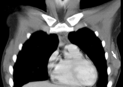
Fig. 3 Chest CT scan showed a well-defined, highly-enhancing mass in the left breast.
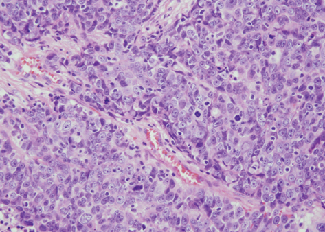
Fig. 4 Photomicrograph of a histopathologic specimen showed small nests of large tumor cells with faintly granular cytoplasm separated by dense collagen bundles (Hematoxylin-eosin, magnification ×200).
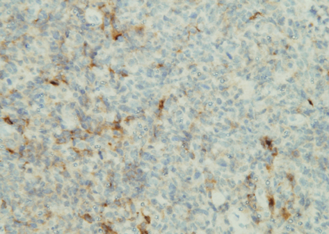
Fig. 5 The tumor stained positive for chromogranin A (magnification ×200) and also expressed neuron-specific enolase and synaptophysin (not shown).
Cited by 1 articles
-
Primary Neuroendocrine Carcinoma of the Breast with Clinical Features of Inflammatory Breast Carcinoma: A Case Report and Literature Review
Do Hyung Lee, Ah Young Park, Bo Kyoung Seo, Young Sik Kim, Ki Yeol Lee, Sang Hoon Cha
J Breast Cancer. 2015;18(4):404-408. doi: 10.4048/jbc.2015.18.4.404.
Reference
-
1. Maluf HM, Koerner FC. Carcinomas of the breast with endocrine differentiation: a review. Virchows Arch. 1994. 425:449–457.
Article2. Papotti M, Macri L, Finzi G, Capella C, Eusebi V, Bussolati G. Neuroendocrine differentiation in carcinomas of the breast: a study of 51 cases. Semin Diagn Pathol. 1989. 6:174–188.3. Sapino A, Righi L, Cassoni P, Papotti M, Pietribiasi F, Bussolati G. Expression of the neuroendocrine phenotype in carcinomas of the breast. Semin Diagn Pathol. 2000. 17:127–137.4. Zekioglu O, Erhan Y, Ciris M, Bayramoglu H. Neuroendocrine differentiated carcinomas of the breast: a distinct entity. Breast. 2003. 12:251–257.
Article5. Tsai WC, Yu JC, Lin CK, Hsieh CT. Primary alveolar-type large cell neuroendocrine carcinoma of the breast. Breast J. 2005. 11:487.
Article6. Gunhan-Bilgen I, Zekioglu O, Ustun EE, Memis A, Erhan Y. Neuroendocrine differentiated breast carcinoma: imaging features correlated with clinical and histopathological findings. Eur Radiol. 2003. 13:788–793.
Article7. Miremadi A, Pinder SE, Lee AH, Bell JA, Paish EC, Wencyk P, Elston CW, Nicholson RI, Blamey RW, Robertson JF, Ellis IO. Neuroendocrine differentiation and prognosis in breast adenocarcinoma. Histopathology. 2002. 40:215–222.
Article8. Krivak TC, McBroom JW, Sundborg MJ, Crothers B, Parker MF. Large cell neuroendocrine cervical carcinoma: a report of two cases and review of the literature. Gynecol Oncol. 2001. 82:187–191.
Article9. Mariscal A, Balliu E, Diaz R, Casas JD, Gallart AM. Primary oat cell carcinoma of the breast: imaging features. AJR Am J Roentgenol. 2004. 183:1169–1171.
Article10. Berruti A, Saini A, Leonardo E, Cappia S, Borasio P, Dogliotti L. Management of neuroendocrine differentiated breast carcinoma. Breast. 2004. 13:527–529.
Article
- Full Text Links
-
- Actions
-
Cited
- CITED
-
- Close
- Share
-
- Similar articles
-
- Small-cell neuroendocrine carcinoma of the breast
- Mammographic, Sonographic, and MRI Features of Primary Neuroendocrine Carcinoma of the Breast: A Case Report
- Primary Neuroendocrine Carcinoma of the Breast: A Case Report and Literature Review
- Primary Neuroendocrine Carcinoma in the Breast: A Case Report
- Two Cases of Neuroendocrine Carcinomas of the Stomach: Large Cell Carcinoma and Small Cell Carcinoma
