J Korean Surg Soc.
2012 Feb;82(2):116-119. 10.4174/jkss.2012.82.2.116.
Small-cell neuroendocrine carcinoma of the breast
- Affiliations
-
- 1Department of Radiology, Eulji General Hospital, Eulji University School of Medicine, Seoul, Korea. jkan0831@eulji.ac.kr
- 2Department of Surgery, Eulji General Hospital, Eulji University School of Medicine, Seoul, Korea.
- 3Department of Pathology, Eulji General Hospital, Eulji University School of Medicine, Seoul, Korea.
- KMID: 2212210
- DOI: http://doi.org/10.4174/jkss.2012.82.2.116
Abstract
- A small-cell carcinoma is one of the histologic subtypes of primary neuroendocrine carcinomas of the breast. A small-cell carcinoma is a rare entity of the breast and exhibits similar morphologic features as neuroendocrine tumors of the gastrointestinal tract and lung. We present the imaging and pathologic findings of a primary small-cell neuroendocrine carcinoma of the breast. This is the first report of a primary small-cell carcinoma arising from the breast in Korea.
MeSH Terms
Figure
-
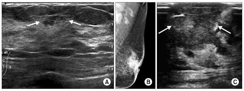
Fig. 1 (A) Sonography showed small irregular hypoechoic lesion with angular margin and spiculations (arrows). (B) Mammography showed ill-defined hyperdense mass in left subareolar area which was adherent to areola. Nipple retraction, diffuse skin thickening, and multiple enlarged lymph nodes in left axilla were noted with shrinkage of volume of left breast. (C) Sonography showed irregular hypoechoic mass with invasion to nipple (arrows).
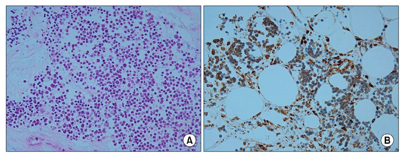
Fig. 2 (A) Microscopic finding showed infiltrating nests of small cells in fibrotic stroma. Tumor cells had small hyperchromatic nuclei and scanty cytoplasms (H&E, ×200). (B) Immunohistochemical stain showed strong positivity of tumor cells for neuron-specific enolase (×400).
Reference
-
1. Sapino A, Bussolati G. Is detection of endocrine cells in breast adenocarcinoma of diagnostic and clinical significance? Histopathology. 2002. 40:211–214.2. Modlin IM, Oberg K, Chung DC, Jensen RT, de Herder WW, Thakker RV, et al. Gastroenteropancreatic neuroendocrine tumours. Lancet Oncol. 2008. 9:61–72.3. Wade PM Jr, Mills SE, Read M, Cloud W, Lambert MJ 3rd, Smith RE. Small cell neuroendocrine (oat cell) carcinoma of the breast. Cancer. 1983. 52:121–125.4. Shin SJ, DeLellis RA, Ying L, Rosen PP. Small cell carcinoma of the breast: a clinicopathologic and immunohistochemical study of nine patients. Am J Surg Pathol. 2000. 24:1231–1238.5. Rosen PP. Rosen PP, editor. Mammary carcinoma with endocrine features. Rosen's breast pathology. 2001. 2nd ed. Philadelphia: Lippincott Williams & Wilkins;503–508.6. Papotti M, Gherardi G, Eusebi V, Pagani A, Bussolati G. Primary oat cell (neuroendocrine) carcinoma of the breast. Report of four cases. Virchows Arch A Pathol Anat Histopathol. 1992. 420:103–108.7. Kinoshita S, Hirano A, Komine K, Kobayashi S, Kyoda S, Takeyama H, et al. Primary small-cell neuroendocrine carcinoma of the breast: report of a case. Surg Today. 2008. 38:734–738.8. Shin SJ, DeLellis RA, Rosen PP. Small cell carcinoma of the breast--additional immunohistochemical studies. Am J Surg Pathol. 2001. 25:831–832.9. Kitakata H, Yasumoto K, Sudo Y, Minato H, Takahashi Y. A case of primary small cell carcinoma of the breast. Breast Cancer. 2007. 14:414–419.10. Mariscal A, Balliu E, Díaz R, Casas JD, Gallart AM. Primary oat cell carcinoma of the breast: imaging features. AJR Am J Roentgenol. 2004. 183:1169–1171.
- Full Text Links
-
- Actions
-
Cited
- CITED
-
- Close
- Share
-
- Similar articles
-
- Primary Small Cell Neuroendocrine Carcinoma of the Breast: A Case Report With Literature Review
- Primary Breast Small Cell Carcinoma With Merkel Cell Features: A Case Report and Literature Review
- Two Cases of Neuroendocrine Carcinomas of the Stomach: Large Cell Carcinoma and Small Cell Carcinoma
- Mammographic, Sonographic, and MRI Features of Primary Neuroendocrine Carcinoma of the Breast: A Case Report
- Primary Neuroendocrine Carcinoma of the Breast: A Case Report and Literature Review
