Relationship between Scanning Laser Polarimetry with Enhanced Corneal Compensation and with Variable Corneal Compensation
- Affiliations
-
- 1Department of Ophthalmology, University of Ulsan, College of Medicine, Asan Medical Center, Seoul, Korea. mskook@amc.seoul.kr
- 2Hangil Eye Hospital, Incheon, Korea.
- KMID: 1107486
- DOI: http://doi.org/10.3341/kjo.2008.22.1.18
Abstract
- PURPOSE: To evaluate the structure-function relationships between retinal sensitivity measured by Humphrey visual field analyzer (HVFA) and the retinal nerve fiber layer (RNFL) thickness measured by scanning laser polarimetry (SLP) with variable corneal compensation (VCC) and enhanced corneal compensation (ECC) in glaucomatous and healthy eyes. METHODS: Fifty-three eyes with an atypical birefringence pattern (ABP) based on SLP-VCC (28 glaucomatous eyes and 25 normal healthy eyes) were enrolled in this cross-sectional study. RNFL thickness was measured by both VCC and ECC techniques, and the visual field was examined by HVFA with 24-2 full-threshold program. The relationships between RNFL measurements in superior and inferior sectors and corresponding retinal mean sensitivity were sought globally and regionally with linear regression analysis in each group. Coefficients of the determination were calculated and compared between VCC and ECC techniques. RESULTS: In eyes with ABP, R2 values for the association between SLP parameters and retinal sensitivity were 0.06-0.16 with VCC, whereas they were 0.21-0.48 with ECC. The association of RNFL thickness with retinal sensitivity was significantly better with ECC than with VCC in 5 out of 8 regression models between SLP parameters and HVF parameters (P<0.05). CONCLUSIONS: The strength of the structure-function association was higher with ECC than with VCC in eyes with ABP, which suggests that the ECC algorithm is a better approach for evaluating the structure-function relationship in eyes with ABP.
MeSH Terms
-
Algorithms
Birefringence
Cornea/physiology
Cross-Sectional Studies
*Diagnostic Techniques, Ophthalmological
Female
Glaucoma/*diagnosis
Humans
Intraocular Pressure
Lasers/diagnostic use
Male
Middle Aged
Nerve Fibers/*pathology
Optic Nerve Diseases/*diagnosis
Prospective Studies
Retinal Ganglion Cells/*pathology
Vision Disorders/*diagnosis
*Visual Fields
Figure
-
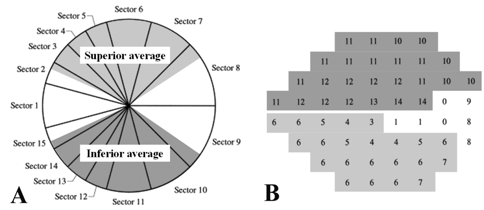
Fig. 1 Correspondence map of the GDx-VCC regional parameters (A) and the HVFA 24-2 paradigm (B) for a right eye. In the present study, peripapillary GDx-VCC measurements and visual field test points were grouped into two regional corresponding sectors of superior and inferior average on the GDx-VCC printouts, based on the article by Garway-Heath et al.21 Corresponding sectors were grayscaled and named after the parameters on the GDx-VCC printouts (Superior and inferior average) in relation to the optic disc.
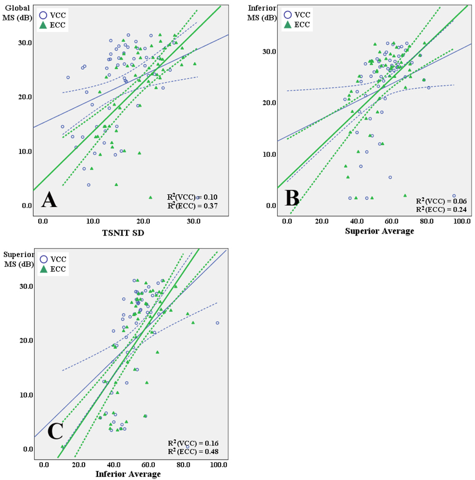
Fig. 2 Scatterplots showing the associations between VCC and ECC parameters and corresponding retinal sensitivities expressed in dB by linear regression in eyes with ABP. (A) TSNIT SD with VCC and ECC versus global mean sensitivity (MS) expressed in dB. (B) Superior average with VCC and ECC versus inferior MS expressed in dB. (C) Inferior average with VCC and ECC versus superior MS expressed in dB. R2 values were significantly higher for linear associations of these SLP parameters with retinal sensitivity with ECC than with VCC (P-values: A, 0.001; B, 0.002; C, <0.001. Wilcoxon signed ranks test of the absolute prediction errors). Solid lines, linear fits; dashed lines, confidence intervals. R2 values are shown for each fit.
Reference
-
1. Reus NJ, Lemij HG. The relationship between standard automated perimetry and GDx-VCC measurements. Invest Ophthalmol Vis Sci. 2004. 45:840–845.2. Lan YW, Henson DB, Kwartz AJ. The correlation between optic nerve head topographic measurements, peripapillary nerve fiber layer thickness, and visual field indices in glaucoma. Br J Ophthalmol. 2003. 87:1135–1141.3. Schlottmann PG, De Cilla S, Greenfield DS, et al. Relationship between visual field sensitivity and retinal nerve fiber layer thickness as measured by scanning laser polarimetry. Invest Ophthalmol Vis Sci. 2004. 45:1823–1829.4. Greenfield DS. Optic nerve and retinal nerve fiber layer analyzers in glaucoma. Curr Opin Ophthalmol. 2002. 13:68–76.5. Blumenthal EZ, Weinreb RN. Assessment of the retinal nerve fiber layer in clinical trials of glaucoma neuroprotection. Surv Ophthalmol. 2001. 45:suppl 3. S305–S312.6. Bagga H, Greenfield DS. Quantitative assessment of structural damage in eyes with localized visual field abnormalities. Am J Ophthalmol. 2004. 137:797–804.7. Zhou Q, Weinreb RN. Individualizd compensation of anterior segment birefringence during scanning laser polarimetry. Invest Ophthalmol Vis Sci. 2002. 43:2221–2228.8. Weinreb RN, Bowd C, Zangwill LM. Glaucoma detection using scanning laser polarimetry with variable corneal polarization compensation. Arch Ophthalmol. 2003. 121:218–224.9. Kook MS, Cho HS, Seong M, Choi J. Scanning laser polarimetry using variable corneal compensation in the detection of glaucoma with localized visual field defects. Ophthalmology. 2005. 112:1970–1978.10. Frenkel S, Slonim E, Horani A, et al. Operator learning effect and interoperator reproducibility of the scanning laser polarimeter with variable corneal compensation. Ophthalmology. 2005. 112:257–261.11. Lleo-Perez A, Ortuno-Soto A, Rahhal MS, et al. Intraobserver reproducibility of retinal nerve fiber layer measurements using scanning laser polarimetry and optical coherence tomography in normal and ocular hypertensive subjects. Eur J Ophthalmol. 2004. 14:523–530.12. Bozkurt B, Irkec M, Gedik S, et al. Effect of peripapillary chorioretinal atrophy on GDx parameters in patients with degenerative myopia. Clin Experiment Ophthalmol. 2002. 30:411–414.13. Tóth M, Holló G. Enhanced corneal compensation for scanning laser polarimetry on eyes with atypical polarisation pattern. Br J Ophthalmol. 2005. 89:1139–1142.14. Hoh ST, Greenfield DS, Liebmann JM, et al. Factors affecting image acquisition during scanning laser polarimetry. Ophthalmic Surg Lasers. 1998. 29:545–551.15. Da Pozzo S, Marchesan R, Canziani T, et al. Atypical pattern of retardation on GDx-VCC and its effect on retinal nerve fiber layer evaluation in glaucomatous eyes. Eye. 2006. 20:769–775.16. Tóth M, Holló G. Evaluation of enhanced corneal compensation in scanning laser polarimetry: comparison with variable corneal compensation on human eyes undergoing LASIK. J Glaucoma. 2006. 15:53–59.17. Reus NJ, Zhou Q, Lemij HG. Enhanced imaging algorithm for scanning laser polarimetry with variable corneal compensation. Invest Ophthalmol Vis Sci. 2006. 47:3870–3877.18. Sehi M, Guaqueta DC, Greenfield DS. An enhancement module to improve the atypical birefringence pattern using scanning laser polarimetry with variable corneal compensation. Br J Ophthalmol. 2006. 90:749–753.19. Bowd C, Tavares IM, Medeiros FA, et al. Retinal Nerve Fiber Layer Thickness and Visual Sensitivity Using Scanning Laser Polarimetry with Variable and Enhanced Corneal Compensation. Ophthalmology. 2007. 144:1259–1265.20. Mai TA, Reus NJ, Lemij HG. Structure function relationship is stronger with enhanced corneal compensation than with variable corneal compensation in scanning laser polarimetry. Invest Ophthalmol Vis Sci. 2007. 48:1651–1658.21. Garway-Heath DF, Poinoosawmy D, Fitzke FW, Hitchings RA. Mapping the visual field to the optic disc in normal tension glaucoma eyes. Ophthalmology. 2000. 107:1809–1815.22. Choi J, Cho HS, Lee CH, Kook MS. Scanning laser polarimetry with variable corneal compensation in the area of apparently normal hemifield in eyes with normal tension glaucoma. Ophthalmology. 2006. 113:1954–1960.23. Reus NJ, Lemij HG. Relationships between standard automated perimetry, HRT confocal scanning laser ophthalmoscopy, and GDx-VCC scanning laser polarimetry. Invest Ophthalmol Vis Sci. 2005. 46:4182–4188.24. Bowd C, Zangwill LM, Weinreb RN. Association between scanning laser polarimetry measurements using variable corneal polarization compensation and visual field sensitivity in glaucomatous eyes. Arch Ophthalmol. 2003. 121:961–966.25. Fedeiros FA, Zangwill LM, Bowd C, Weinreb RN. Comparison of the GDx-VCC scanning laser polarimeter, HRT II confocal scanning laser ophthalmoscope, and StratusOCT optical coherence tomography for the detection of glaucoma. Arch Ophthalmol. 2004. 122:827–837.
- Full Text Links
-
- Actions
-
Cited
- CITED
-
- Close
- Share
-
- Similar articles
-
- Discriminating Ability of Scanning Laser Polarimetry with Variable Corneal Compensation in Normal and Glaucomatous Eyes
- Diagnostic Ability of Scanning Laser Polarimetry with Enhanced Corneal Compensation in the Eye with Typical and Atypical Retadation Pattern
- Scanning Laser Polarimetry Using Variable Corneal Compensation in Detection of Localized Visual Field Defects
- GDx-VCC Performance to Discriminate Normal, Pre-perimetric Glaucomatous Eyes
- Changes in RNFL Thickness According to the Myopia in Patients with Glaucoma and Ocular Hypertension
