Myocardial Bridging of the Left Anterior Descending Coronary Artery: Depiction Rate and Morphologic Features by Dual-Source CT Coronary Angiography
- Affiliations
-
- 1Department of Radiology, Konkuk University Hospital, Konkuk University School of Medicine, Seoul 143-729, Korea. ksm9723@yahoo.co.kr
- 2Department of Thoracic Surgery, Konkuk University Hospital, Konkuk University School of Medicine, Seoul 143-729, Korea.
- KMID: 1102575
- DOI: http://doi.org/10.3348/kjr.2010.11.5.514
Abstract
OBJECTIVE
To evaluate the depiction rate and morphologic features of myocardial bridging (MB) of the left anterior descending coronary artery (LAD) using dual-source CT (DSCT).
MATERIALS AND METHODS
CT scans from a total of 1,353 patients who underwent DSCT were reviewed retrospectively for LAD-MB. Seventy-eight patients were excluded due to poor image quality or poor enhancement of the coronary artery. The length and depth of the MB were analyzed and classified as superficial or deep with respect to the depth (< or = 1 or > 1 mm) of the LAD tunneled segment. Superficial MB was subdivided into complete or incomplete types according to full or partial encasement of the myocardium.
RESULTS
Of the 1,275 patients included in this study, 557 cases of MB were found from 536 patients (42%). Superficial MB was observed in 368 of 557 (66%) cases, and deep MB was seen in 189 of 557 (34%) cases. Superficial MB showed 2 types: complete (128 of 368, 35%) and incomplete (240 of 368, 65%). The mean length of a tunneled segment for superficial MB was 16.4 +/- 8.6 mm. The mean length and depth of a tunneled segment for deep MB were 27.6 +/- 12.8 mm and 3.0 +/- 1.4 mm, respectively. The incidence of atherosclerotic plaques in a 2-cm-long segment proximal to MB was 16%.
CONCLUSION
The depiction rate of LAD-MB using DSCT in a large series of patients was 42%, with two-thirds of MB segments being the superficial type.
Keyword
MeSH Terms
Figure
-
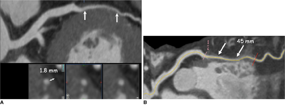
Fig. 1 Depth (A) and length (B) of tunneled segment (arrows) of left anterior descending coronary artery were analyzed on curved multiplanar reformation images using electronic caliper.
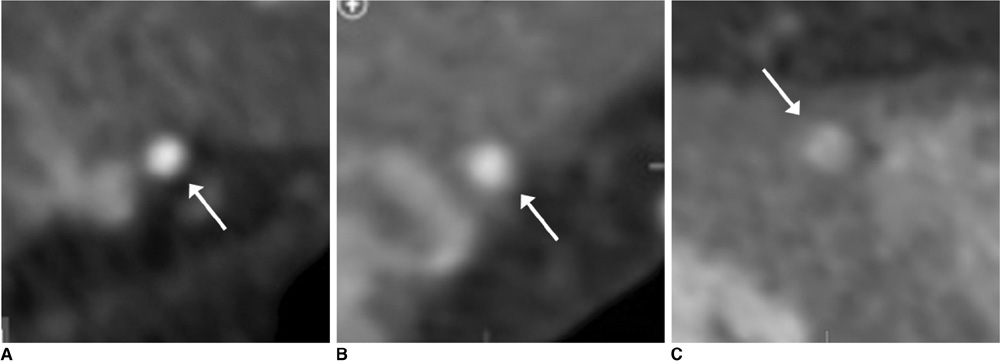
Fig. 2 Myocardial bridging was classified as superficial (A, B) or deep (C) according to depth of tunneled segment (arrows) of left anterior descending coronary artery. Superficial type was subdivided into incomplete (A) and complete (B) types according to extent of vessel encasement by myocardium.
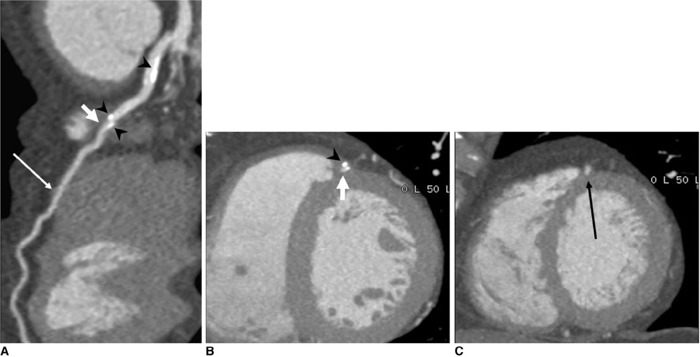
Fig. 3 57-year-old man presented with angina. Curved (A) and short-axis (B, C) multiplanar reformation images showed two tunneled segments (short and long arrows) in mid-left anterior descending coronary artery as well as calcified plaques (arrowheads) in segment proximal to myocardial bridging (A) and in tunneled segment (A, B).

Fig. 4 57-year-old man presented with atypical chest pain. Depth (A) and length (B) of tunneled segment (arrows) of left anterior descending coronary artery were 4.4 mm and 39.3 mm, respectively. Systolic (C) and diastolic (D) curved multiplanar reformation images of tunneled left anterior descending coronary artery segment show that lumen of tunneled segment is compressed by myocardial contraction in systolic phase (C), but had normal diameter in diastolic phase (D). This phenomenon is known as 'milking effect'.
Cited by 1 articles
-
Assessment of Myocardial Bridge by Cardiac CT: Intracoronary Transluminal Attenuation Gradient Derived from Diastolic Phase Predicts Systolic Compression
Mengmeng Yu, Yang Zhang, Yuehua Li, Minghua Li, Wenbin Li, Jiayin Zhang
Korean J Radiol. 2017;18(4):655-663. doi: 10.3348/kjr.2017.18.4.655.
Reference
-
1. Möhlenkamp S, Hort W, Ge J, Erbel R. Update on myocardial bridging. Circulation. 2002. 106:2616–2622.2. Alegria JR, Herrmann J, Holmes DR Jr, Lerman A, Rihal CS. Myocardial bridging. Eur Heart J. 2005. 26:1159–1168.3. Kramer JR, Kitazume H, Proudfit WL, Sones FM Jr. Clinical significance of isolated coronary bridges: benign and frequent condition involving the left anterior descending artery. Am Heart J. 1982. 103:283–288.4. Noble J, Bourassa MG, Petitclerc R, Dyrda I. Myocardial bridging and milking effect of the left anterior descending coronary artery: normal variant or obstruction? Am J Cardiol. 1976. 37:993–999.5. Rossi L, Dander B, Nidasio GP, Arbustini E, Paris B, Vassanelli C, et al. Myocardial bridges and ischemic heart disease. Eur Heart J. 1980. 1:239–245.6. Bourassa MG, Butnaru A, Lespérance J, Tardif JC. Symptomatic myocardial bridges: overview of ischemic mechanisms and current diagnostic and treatment strategies. J Am Coll Cardiol. 2003. 41:351–359.7. Amoroso G, Battolla L, Gemignani C, Panconi M, Petronio AS, Rondine P, et al. Myocardial bridging on left anterior descending coronary artery evaluated by multidetector computed tomography. Int J Cardiol. 2004. 95:335–337.8. Goitein O, Lacomis JM. Myocardial bridging: noninvasive diagnosis with multidetector CT. J Comput Assist Tomogr. 2005. 29:238–240.9. Ko SM, Kim KS. Multidetector-row CT coronary angiographic finding of myocardial bridging. Br J Radiol. 2007. 80:E196–E200.10. Kantarci M, Duran C, Durur I, Alper F, Onbas O, Gulbaran M, et al. Detection of myocardial bridging with ECG-gated MDCT and multiplanar reconstruction. AJR Am J Roentgenol. 2006. 186:S391–S394.11. Zeina AR, Odeh M, Blinder J, Rosenschein U, Barmeir E. Myocardial bridge: evaluation on MDCT. AJR Am J Roentgenol. 2007. 188:1069–1073.12. Konen E, Goitein O, Sternik L, Eshet Y, Shemesh J, Di Segni E. The prevalence and anatomical patterns of intramuscular coronary arteries: a coronary computed tomography angiographic study. J Am Coll Cardiol. 2007. 49:587–593.13. Kawawa Y, Ishikawa Y, Gomi T, Nagamoto M, Terada H, Ishii T, et al. Detection of myocardial bridge and evaluation of its anatomical properties by coronary multislice spiral computed tomography. Eur J Radiol. 2007. 61:130–138.14. Ko SM, Choi JS, Nam CW, Hur SH. Incidence and clinical significance of myocardial bridging with ECG-gated 16-row MDCT coronary angiography. Int J Cardiovasc Imaging. 2008. 24:445–452.15. Leschka S, Koepfli P, Husmann L, Plass A, Vachenauer R, Gaemperli O, et al. Myocardial bridging: depiction rate and morphology at CT coronary angiography--comparison with conventional coronary angiography. Radiology. 2008. 246:754–762.16. Hazirolan T, Canyigit M, Karcaaltincaba M, Dagoglu MG, Akata D, Aytemir K, et al. Myocardial bridging on MDCT. AJR Am J Roentgenol. 2007. 188:1074–1080.17. Konen E, Goitein O, Di Segni E. Myocardial bridging, a common anatomical variant rather than a congenital anomaly. Semin Ultrasound CT MRI. 2008. 29:195–203.18. Jodocy D, Aglan I, Friedrich G, Mallouhi A, Pachinger O, Jaschke W, et al. Left anterior descending coronary artery myocardial bridging by multislice computed tomography: correlation with clinical findings. Eur J Radiol. 2010. 73:89–95.19. Lu GM, Zhang LJ, Guo H, Huang W, Merges RD. Comparison of myocardial bridging by dual-source CT with conventional coronary angiography. Circ J. 2008. 72:1079–1085.20. Kim PJ, Hur G, Kim SY, Namgung J, Hong SW, Kim YH, et al. Frequency of myocardial bridges and dynamic compression of epicardial coronary arteries: a comparison between computed tomography and invasive coronary angiography. Circulation. 2009. 119:1408–1416.21. Canyigit M, Hazirolan T, Karcaaltincaba M, Dagoglu MG, Akata D, Aytemir K, et al. Myocardial bridging as evaluated by 16 row MDCT. Eur J Radiol. 2009. 69:156–164.22. Jacobs JE, Bod J, Kim DC, Hecht EM, Srichai MB. Myocardial bridging: evaluation using single- and dual-source multidetector cardiac computed tomographic angiography. J Comput Assist Tomogr. 2008. 32:242–246.23. La Grutta L, Runza G, Lo Re G, Galia M, Alaimo V, Grassedonio E, et al. Prevalence of myocardial bridging and correlation with coronary atherosclerosis studied with 64-slice CT coronary angiography. Radiol Med. 2009. 114:1024–1036.24. Lubarsky L, Gupta MP, Hecht HS. Evaluation of myocardial bridging of the left anterior descending coronary artery by 64-slice multidetector computed tomographic angiography. Am J Cardiol. 2007. 100:1081–1082.25. Zhang LJ, Yang GF, Zhou CS, Huang W, Lu GM, Shiroishi MS. Multiphase evaluation of myocardial bridging with dual-source computed tomography. Acta Radiol. 2009. 50:775–780.26. Johansen C, Kirsch J, Araoz P, Williamson E. Detection of myocardial bridging by 64-row computed tomography angiography of the coronaries. J Comput Assist Tomogr. 2008. 32:448–451.27. Chen YD, Wu MH, Sheu MH, Chang CY. Myocardial bridging in Taiwan: depiction by multidetector computed tomography coronary angiography. J Formos Med Assoc. 2009. 108:469–474.28. Matt D, Scheffel H, Leschka S, Flohr TG, Marincek B, Kaufmann PA, et al. Dual-source CT coronary angiography: image quality, mean heart rate, and heart rate variability. AJR Am J Roentgenol. 2007. 189:567–573.29. Blankstein R, Shturman LD, Rogers IS, Rocha-Filho JA, Okada DR, Sarwar A, et al. Adenosine-induced stress myocardial perfusion imaging using dual-source cardiac computed tomography. J Am Coll Cardiol. 2009. 54:1072–1084.30. Ko SM, Kim NR, Kim DH, Song MG, Kim JH. Assessment of image quality and radiation dose in prospective ECG-triggered coronary CT angiography compared with retrospective ECG-gated coronary CT angiography. Int J Cardiovasc Imaging. 2010. 26:93–101.31. Baumüller S, Leschka S, Desbiolles L, Stolzmann P, Scheffel H, Seifert B, et al. Dual-source versus 64-section CT coronary angiography at lower heart rates: comparison of accuracy and radiation dose. Radiology. 2009. 253:56–64.32. Donnino R, Jacobs JE, Doshi JV, Hecht EM, Kim DC, Babb JS, et al. Dual-source versus single-source cardiac CT angiography: comparison of diagnostic image quality. AJR Am J Roentgenol. 2009. 192:1051–1056.33. Wang Y, Zhang Z, Kong L, Song L, Merges RD, Chen J, et al. Dual-source CT coronary angiography in patients with atrial fibrillation: comparison with single-source CT. Eur J Radiol. 2008. 68:434–441.34. Meng L, Cui L, Cheng Y, Wu X, Tang Y, Wang Y, et al. Effect of heart rate and coronary calcification on the diagnostic accuracy of the dual-source CT coronary angiography in patients with suspected coronary artery disease. Korean J Radiol. 2009. 10:347–354.35. Brodoefel H, Burgstahler C, Tsiflikas I, Reimann A, Schroeder S, Claussen CD, et al. Dual-source CT: effect of heart rate, heart rate variability, and calcification on image quality and diagnostic accuracy. Radiology. 2008. 247:346–355.36. Bayrak F, Degertekin M, Eroglu E, Guneysu T, Sevinc D, Gemici G, et al. Evaluation of myocardial bridges with 64-slice computed tomography coronary angiography. Acta Cardiol. 2009. 64:341–346.37. Jeong YH, Kang MK, Park SR, Kang YR, Choi HC, Hwang SJ, et al. A head-to-head comparison between 64-slice multidetector computed tomographic and conventional coronary angiographies in measurement of myocardial bridge. Int J Cardiol. 2009. [Epub ahead of print].38. Kim SY, Lee YS, Lee JB, Ryu JK, Choi JY, Chang SG, et al. Evaluation of myocardial bridge with multidetector computed tomography. Circ J. 2010. 74:137–141.39. Atar E, Kornowski R, Fuchs S, Naftali N, Belenky A, Bachar GN. Prevalence of myocardial bridging detected with 64-slice multidetector coronary computed tomography angiography in asymptomatic adults. J Cardiovasc Comput Tomogr. 2007. 1:78–83.
- Full Text Links
-
- Actions
-
Cited
- CITED
-
- Close
- Share
-
- Similar articles
-
- Coronary Flow Velocity Pattern in Patients with Myocardial Bridging of Coronary Artery
- An Overview of Myocardial Bridging With a Focus on Multidetector CT Coronary Angiographic Findings
- Type 4 Dual Left Anterior Descending Artery: A Case Report of a Rare Congenital Coronary Anomaly
- Unusual Coronary Artery Fistula: Left Anterior Descending Coronary Artery - Left Ventricular Fistula Diagnosed by ECG-Gated Multi-Detector Row Coronary CT Angiography
- A Case of Myocardial Bridge in the Left Circumflex Coronary Artery
