Isolated Noncompaction of Ventricular Myocardium: a Magnetic Resonance Imaging Study of 11 Patients
- Affiliations
-
- 1Department of Radiology, Zhongshan Hospital, Fudan University and Shanghai Medical Imaging Institute, 200032 Shanghai, China. zeng-mengsu@zs-hospital.sh.cn
- KMID: 1101922
- DOI: http://doi.org/10.3348/kjr.2011.12.6.686
Abstract
OBJECTIVE
To retrospectively summarize the cardiac magnetic resonance imaging (CMRI) findings of isolated noncompaction of ventricular myocardium (INVM).
MATERIALS AND METHODS
Eleven patients (M:F = 9:2; mean age, 35 years) were evaluated. Steady-state free precession (SSFP), fast spin echo (SE) sequence, SSFP cine imaging, and delayed enhanced inversion recovery spoiled gradient echo (IR-SPGR) sequence were used for showing abnormal myocardium, measuring ratio of noncompacted/compacted myocardium layers (NC/C ratio), and detecting myocardial viability. The left ventricle was divided into nine segments and a NC/C ratio > 2.3 in diastole was used as cutoff value in diagnosing left INVM. The right ventricle was assessed qualitatively.
RESULTS
Cardiac MRI indicated left INVM in seven patients, right INVM in one patient and biventricle INVM in three patients. Characteristic CMRI changes included prominent trabeculations, deep intertrabecular recesses and an increase in the NC/C ratio. The most frequently involved segments was left ventricular apex. Three patients had abnormal high signals within the trabecular structures on SE T2 weighted image. One ventricular aneurysm and one apical thrombus were also observed. Delayed enhancement was seen in six of nine patients with subendocardial and transmural patterns.
CONCLUSION
There are CMRI features that might be characteristic for INVM.
Keyword
MeSH Terms
Figure
-

Fig. 1 Spin echo sequence showing thin epicardial compacted zone and extremely thickened endocardial noncompacted zone in patient with biventricular isolated noncompaction of ventricular myocardium.
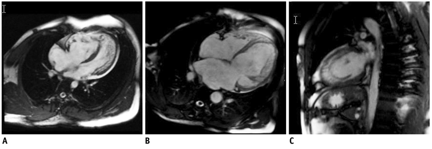
Fig. 2 Steady-state free precession sequence displaying two-layered structure of ventricular myocardium and high signals within trabecular recesses, suggestive of blood flow communicating with ventricular cavity.
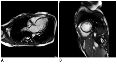
Fig. 3 Steady-state free precession sequence at end of diastolic showing dilated left ventricle and ventricular aneurysm in left ventricular apex (A), septal segment is not affected in short-axis view of left ventricle (B).
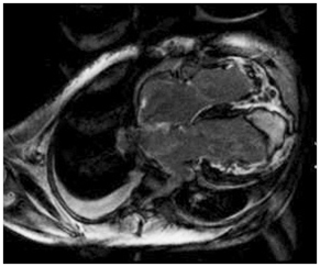
Fig. 4 Apical thrombus (triangle arrow) coexisting with whole heart dilated and delayed enhancement is found in inversion recovery spoiled gradient echo sequence.
Cited by 2 articles
-
Isolated Left Ventricular Noncompaction with a Congenital Aneurysm Presenting with Recurrent Embolism
Jong-Hwa Ahn, Jin-Sin Koh, Jeong Rang Park, Mi Jung Park, Ji Hyun Min, Sang Young Cho, Eun Ju Lee, Wan Chul Kim, Kye Hwan Kim
J Cardiovasc Ultrasound. 2012;20(2):103-107. doi: 10.4250/jcu.2012.20.2.103.Coronary Microembolization with Normal Epicardial Coronary Arteries and No Visible Infarcts on Nitrobluetetrazolium Chloride-Stained Specimens: Evaluation with Cardiac Magnetic Resonance Imaging in a Swine Model
Hang Jin, Hong Yun, Jianying Ma, Zhangwei Chen, Shufu Chang, Mengsu Zeng
Korean J Radiol. 2016;17(1):83-92. doi: 10.3348/kjr.2016.17.1.83.
Reference
-
1. Maron BJ, Towbin JA, Thiene G, Antzelevitch C, Corrado D, Arnett D, et al. Contemporary definitions and classification of the cardiomyopathies: an American Heart Association Scientific Statement from the Council on Clinical Cardiology, Heart Failure and Transplantation Committee; Quality of Care and Outcomes Research and Functional Genomics and Translational Biology Interdisciplinary Working Groups; and Council on Epidemiology and Prevention. Circulation. 2006. 113:1807–1816.2. Chin TK, Perloff JK, Williams RG, Jue K, Mohrmann R. Isolated noncompaction of left ventricular myocardium. A study of eight cases. Circulation. 1990. 82:507–513.3. Dursun M, Agayev A, Nisli K, Ertugrul T, Onur I, Oflaz H, et al. MR imaging features of ventricular noncompaction: emphasis on distribution and pattern of fibrosis. Eur J Radiol. 2010. 74:147–151.4. Petersen SE, Selvanayagam JB, Wiesmann F, Robson MD, Francis JM, Anderson RH, et al. Left ventricular non-compaction: insights from cardiovascular magnetic resonance imaging. J Am Coll Cardiol. 2005. 46:101–105.5. Liu X, Kino A, Francois C, Tuite D, Dill K, Carr JC. Cardiac magnetic resonance imaging findings in a patient with noncompaction of ventricular myocardium. Clin Imaging. 2008. 32:223–226.6. Burke A, Mont E, Kutys R, Virmani R. Left ventricular noncompaction: a pathological study of 14 cases. Hum Pathol. 2005. 36:403–411.7. Ichida F. Left ventricular noncompaction. Circ J. 2009. 73:19–26.8. Attenhofer Jost CH, Connolly HM, Warnes CA, O'Leary P, Tajik AJ, Pellikka PA, et al. Noncompacted myocardium in Ebstein's anomaly: initial description in three patients. J Am Soc Echocardiogr. 2004. 17:677–680.9. Betrian Blasco P, Gallardo Agromayor E. Ebstein's anomaly and left ventricular noncompaction association. Int J Cardiol. 2007. 119:264–265.10. Stollberger C, Blazek G, Winkler-Dworak M, Finsterer J. [Sex differences in left ventricular noncompaction in patients with and without neuromuscular disorders]. Rev Esp Cardiol. 2008. 61:130–136.11. Alhabshan F, Smallhorn JF, Golding F, Musewe N, Freedom RM, Yoo SJ. Extent of myocardial non-compaction: comparison between MRI and echocardiographic evaluation. Pediatr Radiol. 2005. 35:1147–1151.12. Oechslin EN, Attenhofer Jost CH, Rojas JR, Kaufmann PA, Jenni R. Long-term follow-up of 34 adults with isolated left ventricular noncompaction: a distinct cardiomyopathy with poor prognosis. J Am Coll Cardiol. 2000. 36:493–500.13. Sato Y, Matsumoto N, Matsuo S, Kunimasa T, Yoda S, Tani S, et al. Myocardial perfusion abnormality and necrosis in a patient with isolated noncompaction of the ventricular myocardium: evaluation by myocardial perfusion SPECT and magnetic resonance imaging. Int J Cardiol. 2007. 120:e24–e26.14. Hamamichi Y, Ichida F, Hashimoto I, Uese KH, Miyawaki T, Tsukano S, et al. Isolated noncompaction of the ventricular myocardium: ultrafast computed tomography and magnetic resonance imaging. Int J Cardiovasc Imaging. 2001. 17:305–314.15. Finsterer J, Stollberger C, Feichtinger H. Histological appearance of left ventricular hypertrabeculation/noncompaction. Cardiology. 2002. 98:162–164.16. Junga G, Kneifel S, Von Smekal A, Steinert H, Bauersfeld U. Myocardial ischaemia in children with isolated ventricular non-compaction. Eur Heart J. 1999. 20:910–916.17. Jenni R, Wyss CA, Oechslin EN, Kaufmann PA. Isolated ventricular noncompaction is associated with coronary microcirculatory dysfunction. J Am Coll Cardiol. 2002. 39:450–454.18. Jassal DS, Nomura CH, Neilan TG, Holmvang G, Fatima U, Januzzi J, et al. Delayed enhancement cardiac MR imaging in noncompaction of left ventricular myocardium. J Cardiovasc Magn Reson. 2006. 8:489–491.19. Sato Y, Matsumoto N, Yoda S, Inoue F, Kunimoto S, Fukamizu S, et al. Left ventricular aneurysm associated with isolated noncompaction of the ventricular myocardium. Heart Vessels. 2006. 21:192–194.20. Sato Y, Matsumoto N, Matsuo S, Sakai Y, Kunimasa T, Imai S, et al. Right ventricular involvement in a patient with isolated noncompaction of the ventricular myocardium. Cardiovasc Revasc Med. 2007. 8:275–277.21. Fazio G, Lunetta M, Grassedonio E, Gullotti A, Ferro G, Bacarella D, et al. Noncompaction of the right ventricle. Pediatr Cardiol. 2010. 31:576–578.22. Kim KA, Seo JB, Do KH, Heo JN, Lee YK, Song JW, et al. Differentiation of recently infarcted myocardium from chronic myocardial scar: the value of contrast-enhanced SSFP-based cine MR imaging. Korean J Radiol. 2006. 7:14–19.
- Full Text Links
-
- Actions
-
Cited
- CITED
-
- Close
- Share
-
- Similar articles
-
- Noncompaction of Ventricular Myocardium Involving the Right Ventricle
- A case of spongy myocardium initially manifested by ventricular tachycardia in adult
- A case of isolated noncompaction of the ventricular myocardium in an elderly patient
- A Case of Noncompaction of the Ventricular Myocardium Combined with Situs Ambiguous with Polysplenia
- Left Ventricular Noncompaction Associated with Hypertrophic Cardiomyopathy: Morphologic and Functional Evaluation with Multidetector CT
