Atypical Radiological Manifestation of Pulmonary Metastatic Calcification
- Affiliations
-
- 1Department of Internal Medicine, Korea University College of Medicine, Seoul, Korea. keunhae@unitel.co.kr
- 2Department of Pathology, Korea University College of Medicine, Seoul, Korea.
- 3Department of Diagnostic Radiology, Korea University College of Medicine, Seoul, Korea.
- KMID: 1098200
- DOI: http://doi.org/10.3348/kjr.2008.9.2.186
Abstract
- Metastatic pulmonary calcification refers to calcium deposition in the normal pulmonary parenchyma and this deposition is secondary to abnormal calcium metabolism. The most common radiologic manifestation consists of poorly-defined nodular opacities that are mainly seen in the upper lung zone. We present here a case of metastatic pulmonary calcification that manifested as atypical, dense, calcium deposition in airspaces within the previously existing consolidation in the bilateral lower lobes, and this process was accelerated by pneumonia-complicated sepsis in a patient with hypercalcemia that was due to hyperparathyroidism.
MeSH Terms
Figure
-
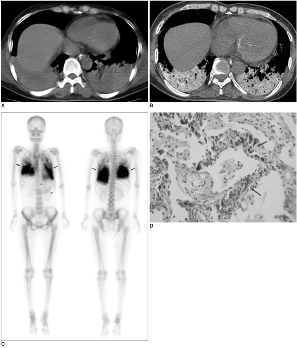
Fig. 1 Metastatic calcification in 48-year-old woman. A. Mediastinal window image of transverse CT scan obtained at level of liver dome and at time of admission shows airspace consolidation in bilateral lower lobes. Note absence of calcification within consolidation at this time. B. Twenty-seven-day-interval follow-up CT scan obtained at level similar to A and after weaning patient from mechanical ventilation demonstrates well-defined bilateral lower lobar consolidation that contains diffuse tissue deposition of calcium, and this is limited to areas of previous consolidation. In addition, scattered calcium deposition was noticed within myocardium. C. Anterior and posterior bone scan images show intense mass-like soft tissue uptake (arrows) of Tc-99m MDP in both lower lung zones and diffusely increased uptakes along stomach wall (arrowheads) without any abnormal bone uptake. D. Alveolar walls are deposited with basophilic spicules of calcium (arrows). Note fibrous tissue within alveolar spaces (Hematoxylin & Eosin staining, ×400).
Reference
-
1. Hartman TE, Müller NL, Primack SL, Johkoh T, Takeuchi N, Ikezoe J, et al. Metastatic pulmonary calcification in patients with hypercalcemia: findings on chest radiographs and CT scans. AJR Am J Roentgenol. 1994. 162:799–802.2. Chung MJ, Lee KS, Franquet T, Müller NL, Han J, Kwon OJ. Metabolic lung disease: imaging and histopathologic findings. Eur J Radiol. 2005. 54:233–245.3. Bendayan D, Barziv Y, Kramer MR. Pulmonary calcifications: a review. Respir Med. 2000. 94:190–193.4. Greenberg S, Suster B. Metastatic pulmonary calcification: appearance on high resolution CT. J Comput Assist Tomogr. 1994. 18:497–499.5. Chan ED, Morales DV, Welsh CH, McDermott MT, Schwarz MI. Calcium deposition with or without bone formation in the lung. Am J Respir Crit Care Med. 2002. 15:1654–1669.6. Kempter H, Hagner G, Savaser AN, Huben H, Minguillon C. Metastatic pulmonary calcification in a patient with nonsecretory multiple myeloma. Respiration. 1986. 49:77–80.7. Neff M, Yalcin S, Gupta S, Berger H. Extensive metastatic calcification of the lung in an azotemic patient. Am J Med. 1974. 56:103–109.8. West JB. Regional differences in blood flow and ventilation in the lung. 1966. Baltimore: Williams and Wilkins;198–254.9. Lingam RK, Teh J, Sharma A, Friedman E. Case report. Metastatic pulmonary calcification in renal failure: a new HRCT pattern. Br J Radiol. 2002. 75:74–77.10. Cotran RS, Kumar V, Robbins SL. Cellular injury and cellular death: pathologic basis of disease. 1994. Philadelphia: W.B.Saunders;1–34.
- Full Text Links
-
- Actions
-
Cited
- CITED
-
- Close
- Share
-
- Similar articles
-
- Metastatic pulmonalry calcification associated with malignant lymphoma: a case report
- Pleural Calcification as a Manifestation of Paragonimiasis: A Report of Two Cases
- Radiological Features of Soft Tissue Calcification: A Pictorial Essay
- Atypical manifestation of solid and papillary epithelial neoplasm of the pancrease: case report
- The radiological evaluation of pulmonary metastases from gastric carcinoma
