Assessment of Left Ventricular Function and Volume in Patients Undergoing 128-Slice Coronary CT Angiography with ECG-Based Maximum Tube Current Modulation: a Comparison with Echocardiography
- Affiliations
-
- 1Department of Cardiology, Kim Hae Jungang Hospital, Gyeongsangnam-do 621-921, Korea.
- 2Department of Radiology, Medical Research Institute, Pusan National University Yangsan Hospital, Pusan National University, School of Medicine, Gyeongsangnam-do 626-770, Korea. kschoo0618@naver.com
- 3Department of Cardiology, Medical Research Institute, Pusan National University Yangsan Hospital, Pusan National University, School of Medicine, Gyeongsangnam-do 626-787, Korea.
- 4Department of Family Medicine, Pusan National University Yangsan Hospital, Pusan National University School of Medicine, Gyeongsangnam-do 626-770, Korea.
- KMID: 1088559
- DOI: http://doi.org/10.3348/kjr.2011.12.2.156
Abstract
OBJECTIVE
To compare multi-detector CT (MDCT) using 128-slice coronary CT angiography (Definition AS+, Siemens Medical Solution, Forchheim, Germany) with ECG-based maximum tube current modulation with echocardiography for the determination of left ventricular ejection fraction (LVEF), end-diastolic volume (EDV), end-systolic volume (ESV), as well as assessing coronary artery image quality and patient radiation dose.
MATERIALS AND METHODS
Thirty consecutive patients (M:F = 20:10; mean age, 57.9 +/- 11.4 years) were referred for MDCT for evaluation of atypical chest pain. EF, EDV and ESV were determined for both MDCT and echocardiography, and the correlation coefficients were assessed. Coronary artery segment subjective image quality (1, excellent; 4, poor) and radiation dose were recorded.
RESULTS
Left ventricular EF, EDV, and ESV were calculated by MDCT and echocardiography and the comparison showed a significant correlation with those estimated by echocardiography (p < 0.05). Consistently, the LVEFs calculated by MDCT and echocardiography were not statistically different. However, LV, EDV and ESV from MDCT were statistically higher than those from echocardiography (p < 0.05). The average image quality score of the coronary artery segment was 1.10 and the mean patient radiation dose was 3.99 +/- 1.85 mSv.
CONCLUSION
Although LV volume was overestimated by MDCT, MDCT provides comparable results to echocardiography for LVEF and LVV, with a low radiation dose.
MeSH Terms
-
Coronary Angiography/*methods
Coronary Disease/*radiography/ultrasonography
Diastole
Echocardiography
*Electrocardiography
Female
Humans
Linear Models
Male
Middle Aged
Radiation Dosage
Radiographic Image Interpretation, Computer-Assisted
Stroke Volume
Systole
*Tomography, X-Ray Computed
Ventricular Dysfunction, Left/*radiography/ultrasonography
Figure
-
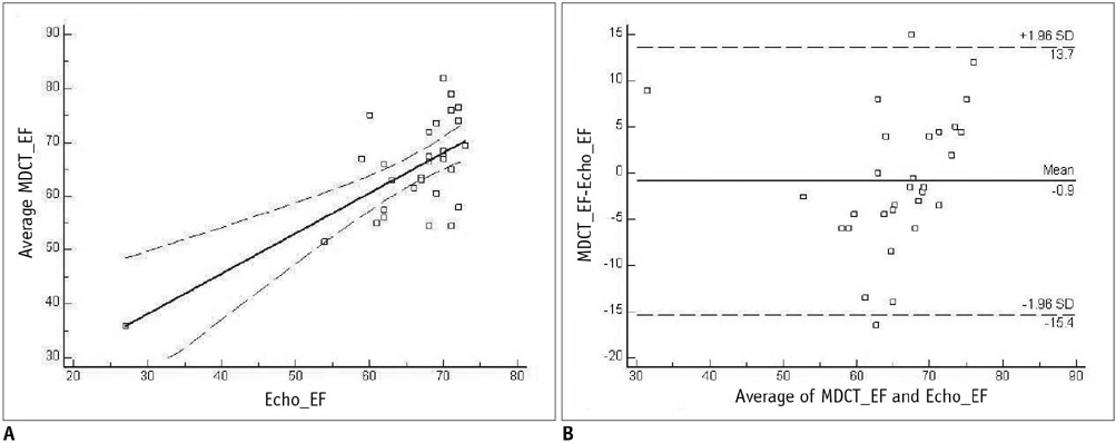
Fig. 1 Comparison of multi-detector CT (MDCT) and two-dimensional echocardiography in assessment of left ventricular ejection fraction. A. Linear regression plot shows correlation between left ventricular ejection fraction (EF) calculated by 128-slice multidetector CT and two-dimensional echocardiography. B. Bland-Altman plot of left ventricular ejection fraction shows difference between each pair plotted against average value of same pair and mean value of differences ± 2 standard deviations (SDs).

Fig. 2 Comparison of multi-detector CT (MDCT) and two-dimensional echocardiography in assessment of left ventricular end-diastolic volume. A. Linear regression plot shows correlation between left ventricular end-diastolic volume (EDV) calculated by 128-slice multidetector CT and two-dimensional echocardiography. B. Bland-Altman plot of left ventricular end-diastolic volume shows difference between each pair plotted against average value of same pair and mean value of differences ± 2 standard deviations (SDs).

Fig. 3 Comparison of multi-detector CT (MDCT) and two-dimensional echocardiography in assessment of left ventricular end-systolic volume. A. Linear regression plot shows correlation between left ventricular end-systolic volume (ESV) calculated by 128-slice multidetector CT and two-dimensional echocardiography. B. Bland-Altman plot of left ventricular end-systolic volume shows difference between each pair plotted against average value of same pair and mean value of differences ± 2 standard deviations (SDs).
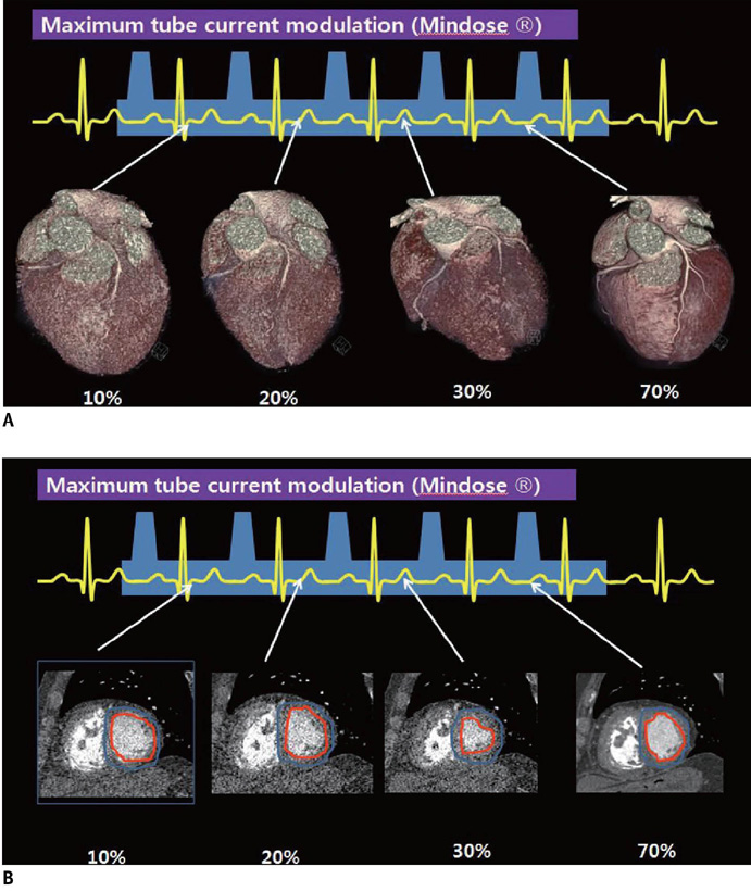
Fig. 4 Difference of image quality depending on exposed maxinum tube current. A. Coronary artery anatomy quality was good at 100% of maximum tube current during mid-diastole and poor outside this phase with 4% of maximum tube current. B. Differentiating endocardium from lumen is available during all cardiac phase including phase with 4% of maximum tube current
Cited by 1 articles
-
Simultaneous Assessment of Left Ventricular Function and Coronary Artery Anatomy by Third-generation Dual-source Computed Tomography Using a Low Radiation Dose
Ji Won Lee, Kyung Jin Nam, Jin You Kim, Yeon Joo Jeong, Geewon Lee, So Min Park, Soo Jin Lim, Ki Seok Choo
J Cardiovasc Imaging. 2020;28(1):21-32. doi: 10.4250/jcvi.2019.0066.
Reference
-
1. Juergens KU, Grude M, Maintz D, Fallenberg EM, Wichter T, Heindel W, et al. Multi-detector row CT of left ventricular function with dedicated analysis software versus MR imaging: initial experience. Radiology. 2004. 230:403–410.2. Mahnken AH, Spuentrup E, Niethammer M, Buecker A, Boese J, Wildberger JE, et al. Quantitative and qualitative assessment of left ventricular volume with ECG-gated multislice spiral CT: value of different image reconstruction algorithms in comparison to MRI. Acta Radiol. 2003. 44:604–611.3. Mahnken AH, Koos R, Katoh M, Spuentrup E, Busch P, Wildberger JE, et al. Sixteen-slice spiral CT versus MR imaging for the assessment of left ventricular function in acute myocardial infarction. Eur Radiol. 2005. 15:714–720.4. Dewey M, Muller M, Teige F, Hamm B. Evaluation of a semiautomatic software tool for left ventricular function analysis with 16-slice computed tomography. Eur Radiol. 2006. 16:25–31.5. White HD, Norris RM, Brown MA, Brandt PW, Whitlock RM, Wild CJ. Left ventricular end-systolic volume as the major determinant of survival after recovery from myocardial infarction. Circulation. 1987. 76:44–51.6. Abada HT, Larchez C, Daoud B, Sigal-Cinqualbre A, Paul JF. MDCT of the coronary arteries: feasibility of low-dose CT with ECG-pulsed tube current modulation to reduce radiation dose. AJR Am J Roentgenol. 2006. 186:S387–S390.7. Hsieh J, Londt J, Vass M, Li J, Tang X, Okerlund D. Step-and-shoot data acquisition and reconstruction for cardiac X-ray computed tomography. Med Phys. 2006. 33:4236–4248.8. Jakobs TF, Becker CR, Ohnesorge B, Flohr T, Suess C, Schoepf UJ, et al. Multislice helical CT of the heart with retrospective ECG gating: reduction of radiation exposure by ECG-controlled tube current modulation. Eur Radiol. 2002. 12:1081–1086.9. Petersilka M, Bruder H, Krauss B, Stierstorfer K, Flohr TG. Technical principles of dual source CT. Eur J Radiol. 2008. 68:362–368.10. Schiller NB, Shah PM, Crawford M, DeMaria A, Devereux R, Feigenbaum H, et al. American Society of Echocardiography Committee on Standards. Subcommittee on Quantitation of Two-Dimensional Echocardiograms. Recommendations for quantitation of the left ventricle by two-dimensional echocardiography. J Am Soc Echocardiogr. 1989. 2:358–336.11. Christner JA, Kofler JM, McCollough CH. Estimating effective dose for CT using dose-length product compared with using organ doses: consequences of adopting International Commission on Radiological Protection publication 103 or dual-energy scanning. AJR Am J Roentgenol. 2010. 194:881–889.12. Raff GL, Gallagher MJ, O'Neill WW, Goldstein JA. Diagnostic accuracy of noninvasive coronary angiography using 64-slice spiral computed tomography. J Am Coll Cardiol. 2005. 46:552–557.13. Hoffmann U, Moselewski F, Cury RC, Ferencik M, Jang IK, Diaz LJ, et al. Predictive value of 16-slice multidetector spiral computed tomography to detect significant obstructive coronary artery disease in patients at high risk for coronary artery disease: patient-versus segment-based analysis. Circulation. 2004. 110:2638–2643.14. Butler J, Shapiro MD, Jassal DS, Neilan TG, Nichols J, Ferencik M, et al. Comparison of multidetector computed tomography and two-dimensional transthoracic echocardiography for left ventricular assessment in patients with heart failure. Am J Cardiol. 2007. 99:247–249.15. Hausleiter J, Meyer T, Hermann F, Hadamitzky M, Krebs M, Gerber TC, et al. Estimated radiation dose associated with cardiac CT angiography. JAMA. 2009. 301:500–507.16. de Graaf FR, Schuijf JD, van Velzen JE, Nucifora G, Kroft LJ, de Roos A, et al. Assessment of global left ventricular function and volumes with 320-row multidetector computed tomography: a comparison with 2D-echocardiography. J Nucl Cardiol. 2010. 17:225–231.
- Full Text Links
-
- Actions
-
Cited
- CITED
-
- Close
- Share
-
- Similar articles
-
- Comparison of Left and Right Ventricular Volume and Cardiac Output by MRI and Echocardiography
- Myocardial Contractility, Perfusion, and Viability Analysis Using Multidetector CT in Patients with Ischemic Heart Disease
- Unusual Coronary Artery Fistula: Left Anterior Descending Coronary Artery - Left Ventricular Fistula Diagnosed by ECG-Gated Multi-Detector Row Coronary CT Angiography
- Noninvasive Detection of Coronary Atherosclerotic Plaques and Assessment of Stenosis Degree at Multidetector CT Coronary Angiography
- Coincident Takotsubo Cardiomyopathy and Coronary Artery Disease
