J Vet Sci.
2011 Sep;12(3):295-297. 10.4142/jvs.2011.12.3.295.
Development of a novel diagnostic test for detection of bovine viral diarrhea persistently infected animals using hair
- Affiliations
-
- 1Veterinary Diagnostic Laboratory, College of Veterinary Medicine, University of Illinois, Urbana, IL 61802, USA. ksingh08@illinois.edu
- 2Veterinary Teaching Hospital, College of Veterinary Medicine, University of Illinois, Urbana, IL 61802, USA.
- 3Department of Veterinary Science, Wyoming State Veterinary Laboratory, Laramie, WY 82070, USA.
- KMID: 1067404
- DOI: http://doi.org/10.4142/jvs.2011.12.3.295
Abstract
- The purpose of this study was to determine whether manually plucked hairs might serve as an alternative sample for a quantitative real time polymerase chain reaction (qRT-PCR) testing. Twenty three, 1~3 week old, non-bovine viral diarrhea virus (BVDV) vaccinated calves, found to be positive for BVDV by immunohistochemical staining, were selected and hairs were manually plucked from the ear. qRT-PCR was performed on samples consisting of more than 30 hairs (30~100) and whole blood. All 23 animals were positive for the virus by qRT-PCR performed on the whole blood and when samples of more than 30 hairs were assayed. Additionally, qRT-PCR was performed on groups of 10 and 20 hairs harvested from 7 out of 23 immunohistochemical staining-positive calves. When groups of 20 and 10 hairs were tested, 6 and 4 animals, respectively, were positive for the virus.
MeSH Terms
-
Animals
Antibodies, Viral/analysis/diagnostic use
Bovine Virus Diarrhea-Mucosal Disease/blood/*diagnosis/virology
Cattle
Diarrhea Virus 1, Bovine Viral/genetics/*isolation & purification
Diarrhea Virus 2, Bovine Viral/genetics/*isolation & purification
Hair/virology
RNA, Viral/chemistry/genetics
Real-Time Polymerase Chain Reaction/methods/*veterinary
Figure
-
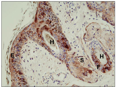
Fig. 1 Multiple hair follicular cells were positive for bovine viral diarrhea virus (BVDV) antigen using immunohistochemistry. Note the positive staining in epidermal cells (E), hair (H), follicular cells (F), and sebaceous glands (S).
Reference
-
1. Bhudevi B, Weinstock D. Fluorogenic RT-PCR assay (TaqMan) for detection and classification of bovine viral diarrhea virus. Vet Microbiol. 2001. 83:1–10.
Article2. Cornish TE, van Olphen AL, Cavender JL, Edwards JM, Jaeger PT, Vieyra LL, Woodard LF, Miller DR, O'Toole D. Comparison of ear notch immunohistochemistry, ear notch antigen-capture ELISA, and buffy coat virus isolation for detection of calves persistently infected with bovine viral diarrhea virus. J Vet Diagn Invest. 2005. 17:110–117.
Article3. Driskell EA, Ridpath JF. A survey of bovine viral diarrhea virus testing in diagnostic laboratories in the United States from 2004 to 2005. J Vet Diagn Invest. 2006. 18:600–605.
Article4. Reichel MP, Hill FI, Voges H. Does control of bovine viral diarrhoea infection make economic sense? N Z Vet J. 2008. 56:60–66.
Article5. Ridpath JF, Fulton RW. Knowledge gaps impacting the development of bovine viral diarrhea virus control programs in the United States. J Am Vet Med Assoc. 2009. 235:1171–1179.
Article
- Full Text Links
-
- Actions
-
Cited
- CITED
-
- Close
- Share
-
- Similar articles
-
- Tissue distribution of bovine viral diarrhea virus antigens in persistently infected cattle
- Genetic characterization of bovine viral diarrhea virus strains in Beijing, China and innate immune responses of peripheral blood mononuclear cells in persistently infected dairy cattle
- Prevalence of bovine viral diarrhea virus from Korean native cattle farms in Jeju
- Genetic Typing of Bovine Viral Diarrhea Viruses (BVDV) Circulating in Korea
- Klebsiella pneumoniae infection secondary to bovine viral diarrhea in two prematurely born calves
